 Clinical suspicion of abdominal aortic aneurysm (AAA)
 Unexplained abdominal, back, or flank pain, particularly in the older patient
 Unexplained hypotension
 Syncope in the setting of abdominal pain
 Palpable and/or pulsatile abdominal mass
CONTRAINDICATIONS
 None: No contrast or radiation involved
LANDMARKS
 The proximal aorta is located in the subxiphoid area
 The aorta bifurcates into the iliac vessels at the level of the umbilicus
 On the ultrasound display (FIGURE 12.1), first locate the vertebral body. The transverse proximal aorta is located above (anterior to) the vertebral body, to the right of the inferior vena cava (IVC), and below (posterior to) the superior mesenteric artery
TECHNIQUE
 Apply ultrasond gel on the patient’s abdomen, from the xiphoid process to just distal to the umbilicus
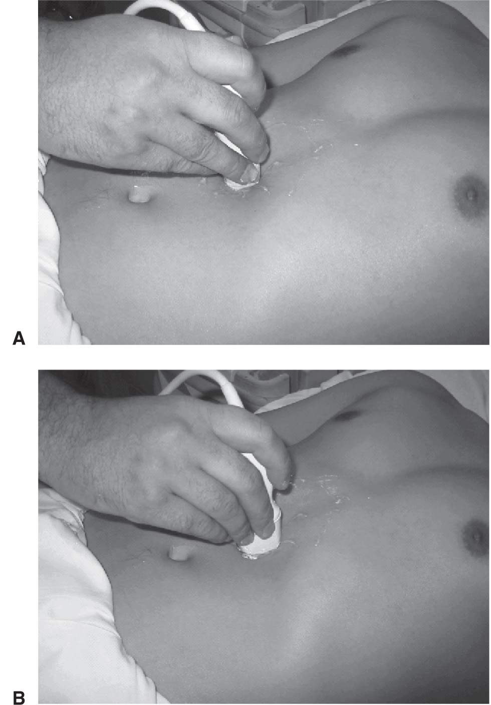
 Using a standard 3.5- to 5.0-MHz probe with the selection marker to the patient’s right, identify the proximal aorta in the epigastric area (FIGURE 12.2)
 Once the aorta is identified, scan the entire length of the vessel to the iliac bifurcation at the umbilicus
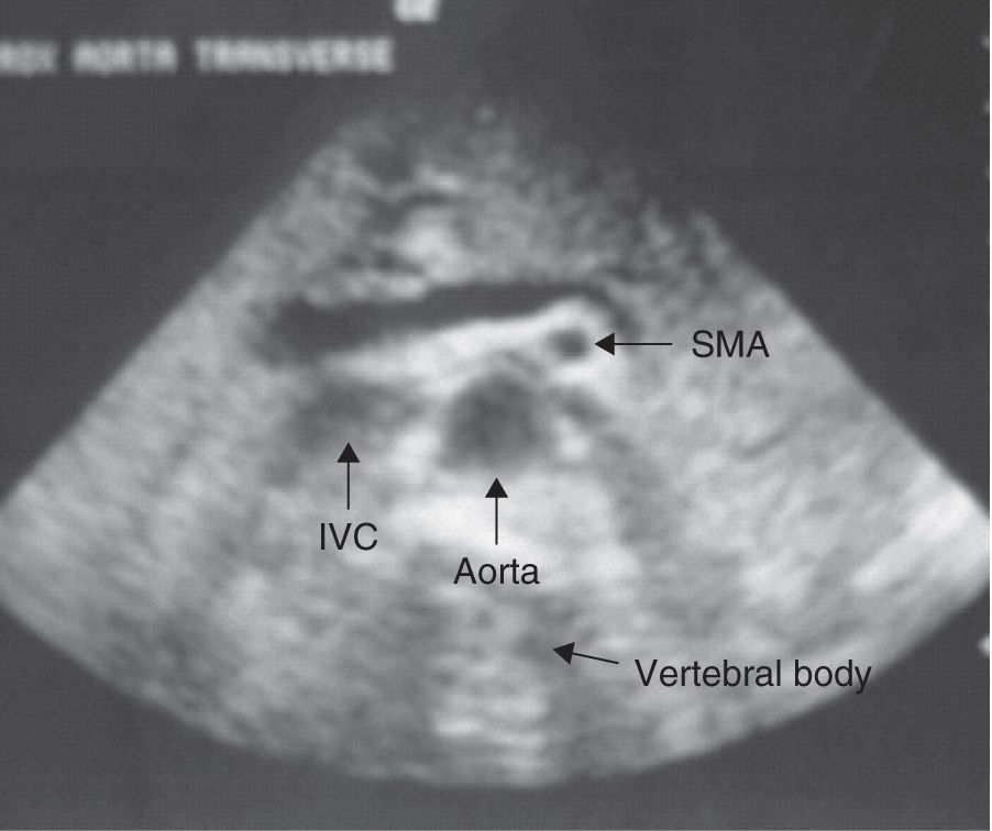
FIGURE 12.1 Transverse proximal aorta anatomy. IVC, inferior vena cava; SMA, superior mesenteric artery. (Courtesy of David Riley, MD.)
 Measure the vessel diameter from outer wall to outer wall to avoid underestimation
 Vessel measurement in the following views permits adequate screening for AAA (FIGURE 12.3):
 Transverse proximal aorta
 Transverse middle aorta
 Transverse distal aorta
 Transverse view of iliac arteries at the bifurcation
 Longitudinal aorta
 If an aneurysm is detected (TABLE 12.1), its relation to the renal arteries and aortic bifurcation should be documented if possible (i.e., proximal, distal)
 If the aorta is not readily identified, try the following techniques:
 Apply gentle downward pressure with the probe to displace bowel gas
 Increase the depth of penetration on the ultrasound display
 Reimaging after several minutes may permit improved visualization as intestinal peristalsis displaces bowel
 Place the patient in the right or left lateral decubitus position
 Approach para-midline by directing the probe toward the spine to avoid bowel gas
 Image coronally through the liver
 Image inferiorly to the umbilicus and direct the probe cephalad
COMPLICATIONS
 Delay in definitive surgical treatment in order to perform study. Computed tomography (CT) evaluation typically poses a greater time delay.
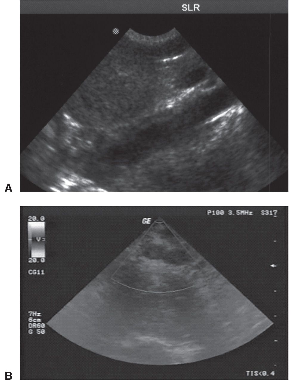
FIGURE 12.3 A: Longitudinal aorta. B: Transverse image of the common iliac bifurcation. (Images courtesy of David Riley, MD.)



Full access? Get Clinical Tree


