 Yes or No: Is there intra-abdominal fluid or fluid around the heart?
 The assessment of blunt thoracoabdominal trauma with significant mechanism of injury
 The assessment of penetrating torso trauma if operative management is not immediately indicated
CONTRAINDICATIONS
 The FAST examination should never delay a patient’s transport to the operating room when operative management is clearly indicated
RISKS/CONSENT ISSUES
 The only theoretical risk is allergy to the ultrasound gel
ADVANTAGES
 Noninvasive and no sedation required
 Can be performed at the bedside while resuscitative efforts are simultaneously being performed
 Can be performed at the bedside on patients too unstable for the computed tomographic imaging suite
 Can be repeated serially along with changes in symptoms or hemodynamic stability
LANDMARKS
 Subcostal
 Probe placed in the subxiphoid region pointed toward the heart detects fluid in pericardial sac
 Hepatorenal
 Probe placed in the right midaxillary line between the 8th and 11th ribs detects fluid in the hepatorenal space (Morison pouch)
 Splenorenal
 Probe placed in the left posterior axillary line between the 8th and 11th ribs detects fluid in the splenorenal recess
 Suprapubic
 Probe placed 2 cm superior to the symphysis pubis detects fluid in the retrovesical or retrouterine space
TECHNIQUE
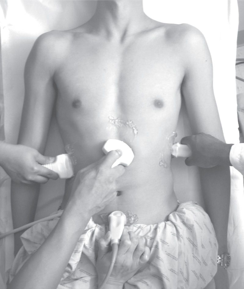
The standard four FAST views: Subcostal, hepatorenal pouch (Morison), splenorenal, and suprapubic (FIGURE 18.1)
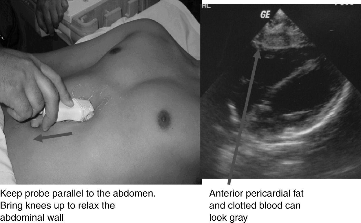
 Subcostal/Subxiphoid View (FIGURE 18.2)
 With the probe in the transverse plane, place it in the subxiphoid area and aim toward the patient’s left shoulder to see a four-chambered view of the heart
 Sweep the probe anteriorly and posteriorly to view the entire pericardium
 Unclotted blood will appear as an anechoic black “stripe” within the hyperechoic pericardial sac (FIGURE 18.3)



Full access? Get Clinical Tree


