 Identify and differentiate cellulitis from abscess with overlying cellulitis
 Procedural guidance for incision and drainage of abscess
 Assess soft-tissue masses
CONTRAINDICATIONS
 None
None
RISKS/CONSENT ISSUES
 Contact dermatitis from ultrasound gel (very rare)
TECHNIQUE
 Linear transducer (7.5–10+ MHz)
 Sonographic grip—always have part of the hand, i.e., the little finger, resting upon the body of the patient when possible, as this helps stabilize the probe for fine movements
 Reduce depth
 Adjust gain using a fluid-filled structure as the standard (anechoic fluid or blood should be crisply anechoic)
 Caliper function can be used to measure abscesses in two or three planes
 Assess for vascularity using color Doppler, as the presence of vascularity may result in a contraindication to incision and drainage
NORMAL SKIN AND SOFT TISSUE (FIGURE 88.1)
 Adjust depth to focus only on region of interest, typically 2 to 4 cm
 Scan a wide region, starting away from the region of interest to assess presumably normal skin and soft tissue
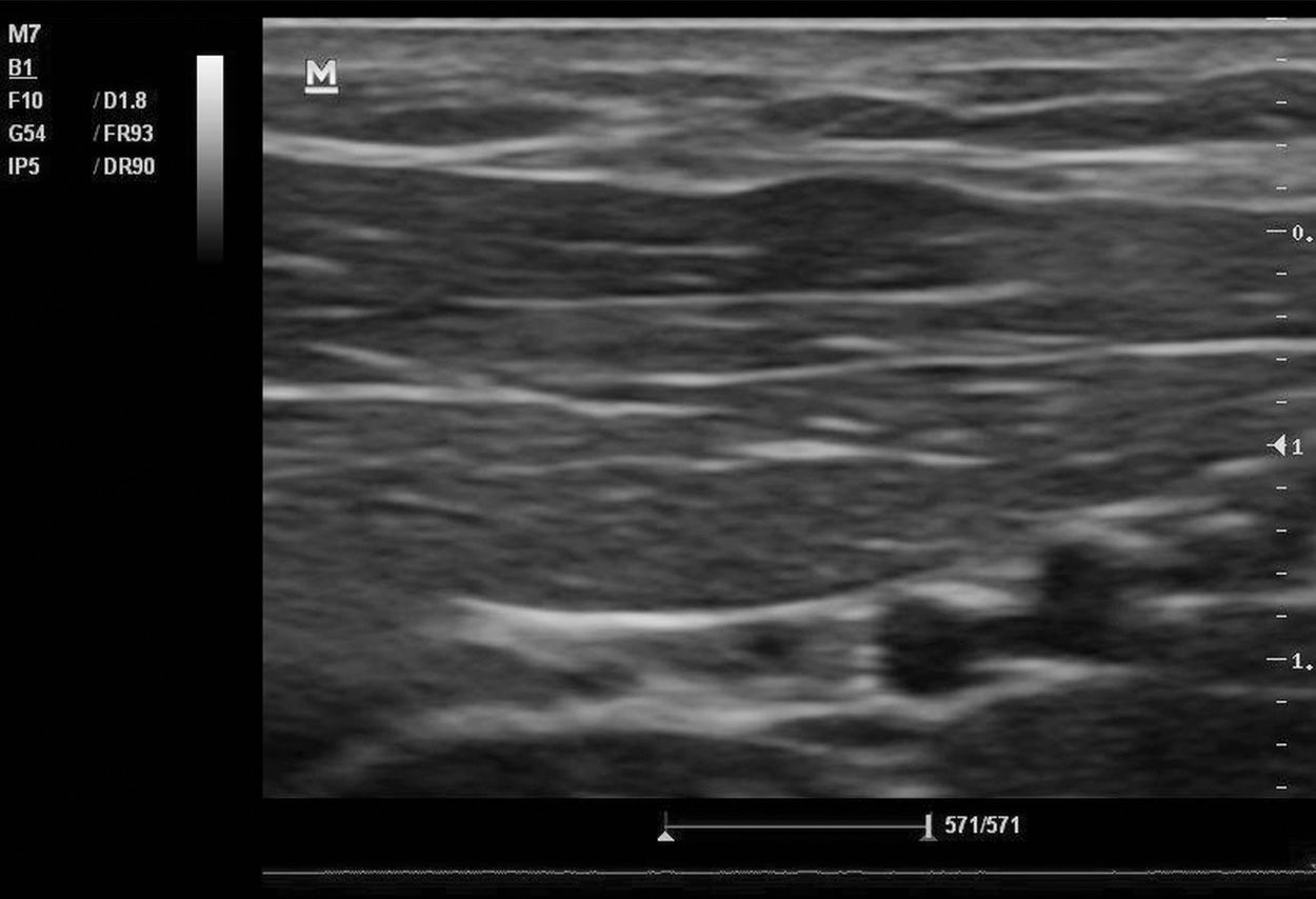
 Be familiar with the appearance of soft tissue and the structures within it (skin, subcutaneous tissue, blood vessels, lymph node, nerve, muscle fascicles, tendon, bone)
PATHOLOGY
 Cellulitis (FIGURE 88.2)
 Diffuse thickened (slightly hyperechoic) subcutaneous layer and fluid (anechoic) dissecting the deeper layers of the skin and fat, creating the pathognomonic cobblestone appearance
 Can see thin hyperechogenic transverse layers between layers of normal dermis
 Posterior acoustic enhancement
 Abscess (FIGURES 88.3–88.5)
 Variable appearance on ultrasound
 Typically see well-defined border, but possibly irregular
 May see anechoic fluid-filled pocket with or without septae supporting loculation (debris)
 May see posterior acoustic enhancement (shadowing)



Full access? Get Clinical Tree


