 Clinical suspicion
 Swollen/red peritonsillar region causing uvular shift
 Fluctuance of area
 Interim treatment for peritonsillar closed space infection until tonsillectomy
CONTRAINDICATIONS
 Extension into the deep neck tissue
 Septicemia/toxic appearance
 Airway obstruction
 Severe trismus
 Coagulopathy
 General Basic Steps
 Patient preparation
 Analgesia
 Visualization
 Needle aspiration/incision and drainage (I&D)
LANDMARKS
 Superior lateral border of affected tonsil, or area of most fluctuance
 Aspirate peritonsillar abscess’ (PTA’s) superior pole first, then middle pole, and finally the inferior pole (FIGURE 76.1)
TECHNIQUE
 Patient Preparation
 Cooperative patient sitting upright in a chair with occipital support
 Consider intravenous analgesia or sedation
 Digital exam key: Must feel abscess!
 Use ultrasound (endocavitary probe) to assess volume, location, and relationship to the carotid artery (FIGURE 76.2)
 Needle Aspiration
 Anesthetize with benzocaine spray or have patient gargle viscous lidocaine
 Have patient depress own tongue by holding laryngoscope, insert as you would for intubation. Patient will be less likely to trigger own gag reflex while pulling down on blade (FIGURE 76.3)
 Anesthetize locally with 1 to 2 mL of 1% lidocaine via 27-gauge needle
 Use a long spinal needle so visualization is not obscured by syringe
 Cut the distal 1 cm off of the needle cover and recap the needle, thereby preventing the needle from penetrating >1 cm (FIGURE 76.4)
 Insert spinal needle at area of greatest fluctuance (usually the superior pole) and aspirate the pus
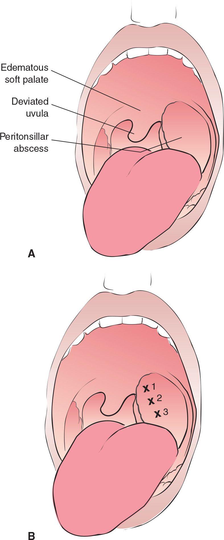
FIGURE 76.1 A: Peritonsillar abscess. The tonsil is displaced forward and inferomedial, the uvula is deviated toward the unaffected tonsil, and the soft palate is edematous and ruborous. B: Recommended sites for three-point needle aspiration of a peritonsillar abscess. (From Saladino RA. Pharyngeal procedures. In: Henretig FM, King C, eds. Textbook of Pediatric Emergency Procedures. Philadelphia, PA: Williams & Wilkins; 1997:692, 696, with permission.)



Full access? Get Clinical Tree


