Shoulder Dislocation and Reduction
Olabiyi Akala and Maureen Gang
INDICATIONS
 History and clinical examination consistent with shoulder dislocation
 Anterior Dislocation (~95%)
 Mechanism
 Force applied to an externally rotated, abducted, and extended arm
 Rarely secondary to a blow to the posterior shoulder
 Examination
 Prominent humeral head anteriorly and a shallow depression inferior to the acromion may be observed
 Affected extremity usually held in abduction and external rotation
 Posterior Dislocation (2%–4%)
 Mechanism
 Axial loading of adducted and internally rotated arm
 Less commonly due to direct blow to anterior shoulder or fall on an outstretched arm
 May result from violent muscle contractions: e.g., seizures, electric shock, psychiatry patients
 Examination
 Prominence of posterior shoulder with flattening anteriorly; may be subtle
 Affected extremity typically held in adduction and internal rotation
 Patient usually unable to externally rotate affected extremity
 Inferior dislocation (luxatio erecta)—rare
 Mechanism
 Forceful hyperabduction of the affected extremity
 Examination
 Affected arm is held above the head
 Patient is unable to adduct the affected extremity
 Radiographs demonstrate glenohumeral dislocation
CONTRAINDICATIONS
 Any associated fracture—particularly fracture of the humeral neck
 Obtain orthopedic consultation
 Any associated neurologic deficit
 Closed reduction may still be attempted but multiple attempts should be avoided
RISKS/CONSENT ISSUES
 Recurrent dislocation
 Risk dependent on age at initial dislocation, with recurrence risk up to 90% for those <20, up to 70% for those between 20 and 40 and between 2% and 4% for those older than 40
 Increased risk of associated rotator cuff injuries in patients >40 years of age
 Complications of reduction
 Risks associated with procedural sedation
 Neurovascular injury
 Fracture of humerus and glenoid
 General Basic Steps
 Thorough examination of affected extremity, including neurovascular status
 Analgesia/sedation/muscle relaxation
 Reduction via preferred technique
 Postreduction care and follow-up
LANDMARKS—FIGURE 62.1
 Technique
 Physical Examination
 Compare both the affected and unaffected extremities
 Perform a thorough neurovascular examination of the injured extremity
 A sensory deficit over the deltoid (the so-called sergeant’s-stripe pattern) or an impaired deltoid contraction implies an axillary nerve injury
 All major nerves to the arm should be assessed as injuries to the brachial plexus, ulnar, and radial nerves have been reported
 Radiographs
 Obtain before reduction if the clinician is unsure of the position/type of dislocation or if there is concern for an associated fracture
 May defer prereduction films if the clinician is confident of an anterior dislocation based on physical examination, the patient is <40, with a history of recurrent dislocations, and the mechanism of the dislocation is not associated with direct trauma
 Anteroposterior (AP), scapular Y, and axillary lateral view should be obtained
 A single x-ray view should never be used to diagnose a shoulder dislocation
 In anterior dislocations, the humeral head is anterior in the axillary view (using the coracoid process as a point of orientation, and anterior to the center of Y in the trans-scapular view
 In posterior dislocations, the AP view may be diagnostic if it shows a partial vacancy of the glenoid fossa (vacant glenoid sign) and >6 mm space between the glenoid rim and humeral head (positive rim sign). The humeral head is posterior on axillary view and posterior to center Y on trans-scapular view.
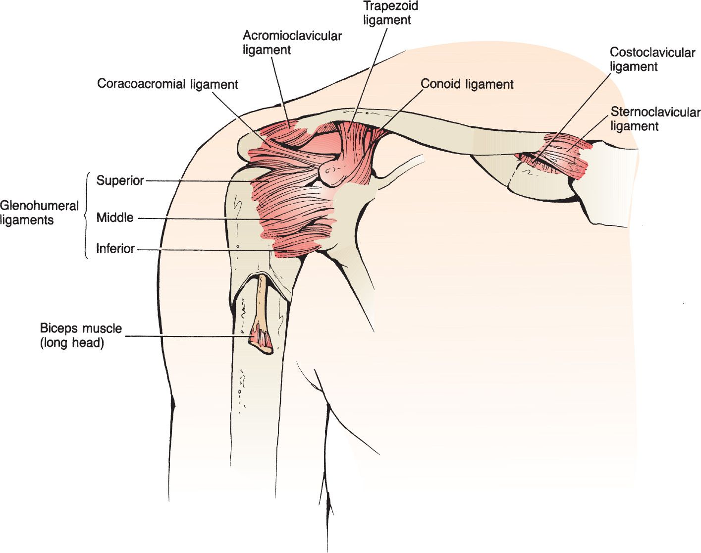
FIGURE 62.1 The essential anatomy of the shoulder. (From Sherman S. Shoulder injuries. In: Wolfson AB, ed. Harwood-Nuss’ Clinical Practice of Emergency Medicine. 6th ed. Philadelphia, PA: Lippincott Williams & Wilkins; 2015:248, with permission.)
 Sedation, Analgesia, and Muscle Relaxation
 Adequate analgesia, muscle relaxation, and/or sedation help facilitate successful reduction
 A recent systematic review of intra-articular lidocaine vs procedural sedation showed no significant difference in reduction success rates, pain during reduction, and pain after reduction
 It is reasonable to attempt initial reduction with intra-articular local anesthetic; if unsuccessful, the clinician may consider procedural sedation for subsequent attempts
 Ensure that the patient relates the use of intra-articular lidocaine to the orthopedic surgeon during follow-up
 Intra-articular Injection of Lidocaine
 Cleanse the shoulder with povidone–iodine solution
 Insert the needle 2 cm inferiorly and directly lateral to the acromion, in the lateral sulcus left by the absent humeral head
 Fill a 20-mL syringe with 1% lidocaine. Attach a 1.5-inch 20-gauge needle to the syringe (FIGURE 62.2).
 Withdraw to ensure you are not in a blood vessel prior to the injection of 15 to 20 mL of lidocaine into the joint space
 Shoulder Reduction
 The guiding principle for all methods of reduction should be a gradual and gentle application of technique (FIGURE 62.3)
 The treating physician should be comfortable with several methods of reduction because no technique is 100% effective. The following techniques are described in this chapter:
 Stimson maneuver
 Scapular manipulation
 Traction–countertraction
 Milch technique
 Hennepin or external rotation method
 Cunningham technique
 Posterior dislocation reduction
 Postreduction Care
 Obtain postreduction x-rays
 Perform a postreduction neurovascular assessment and document the findings
 Position at discharge is controversial. Evidence regarding external rotation splinting is still evolving. Patients should be placed in a shoulder immobilizer or sling and swath for 2 to 3 weeks.
 Arrange orthopedic follow-up in 1 to 2 weeks
 Older patients (<40) should have early follow-up within ~1 week to prevent adhesive capsulitis (frozen shoulder)
 Stimson Maneuver
 Patient is positioned prone with dislocated arm overhanging the bed
 Weight of 5 to 15 lb (initially supported by the physician) is strapped to the wrist of the affected extremity
 Traction is gradually exerted on the shoulder by slow and steady release of the physician’s support
 Up to 30 minutes of sustained, steady traction may be necessary for reduction
 Reduction may be facilitated by delicate external rotation of the affected extremity
 Advantages: Can be performed by the lone practitioner without assistance
 Disadvantages: Often requires more time and materials (weights and straps) than may be readily available (FIGURE 62.4). Not appropriate for all patients, particularly those with respiratory compromise.
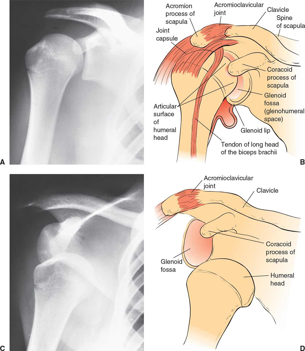
FIGURE 62.2 A, B: Normal shoulder joint. C, D: Anterior dislocation of the shoulder. (From Young GM. Reduction of common joint dislocations and subluxations. In: Henretig FM, King C, eds. Textbook of Pediatric Emergency Procedures. Philadelphia, PA: Williams & Wilkins; 1997:1083, with permission.)



Full access? Get Clinical Tree


