 Inadequate oxygenation or ventilation
 Airway obstruction
 Loss of protective airway reflexes (e.g., depressed cough and gag reflexes)
 Excess work of breathing
 Nonresponsive and apneic
CONTRAINDICATIONS
 Absolute Contraindications
 None for unstable patients (i.e., “crash” airway)
 Relative Contraindications
 In these circumstances one should consider consultation with anesthesiologist/intensivist, alternative techniques, and/or sedation without paralysis
 Infectious: Epiglottitis, croup, retropharyngeal abscess, bacterial tracheitis
 Noninfectious: Anaphylaxis/angioedema, foreign body, trauma, burns
 Congenital anomalies (e.g., cleft palate, micrognathia)
 Unanticipated difficult airway (e.g., multiple failed attempts)
RISKS/CONSENT ISSUES
 Airway trauma
 Arrhythmia (e.g., bradyarrhythmia)
 Aspiration of stomach contents
 Esophageal intubation
 Increase in blood pressure and intracranial pressure (ICP)
 Hypoxemia
 Pain
Pain
LANDMARKS
 Anatomical differences in children (FIGURE 89.1):
 Larger tongue
 Larger and floppy epiglottis
 Narrower cricoid ring
 Larger occiput
 The glottic opening is more cranial and anterior in children and is located at:
 C1 in infancy
 C3–C5 at age 7
 C4–C6 in the adult (Figure 89.1)
 Differences are most pronounced under 2 years, transition from 2 to 8 years, then approach small adult anatomy by 8 years
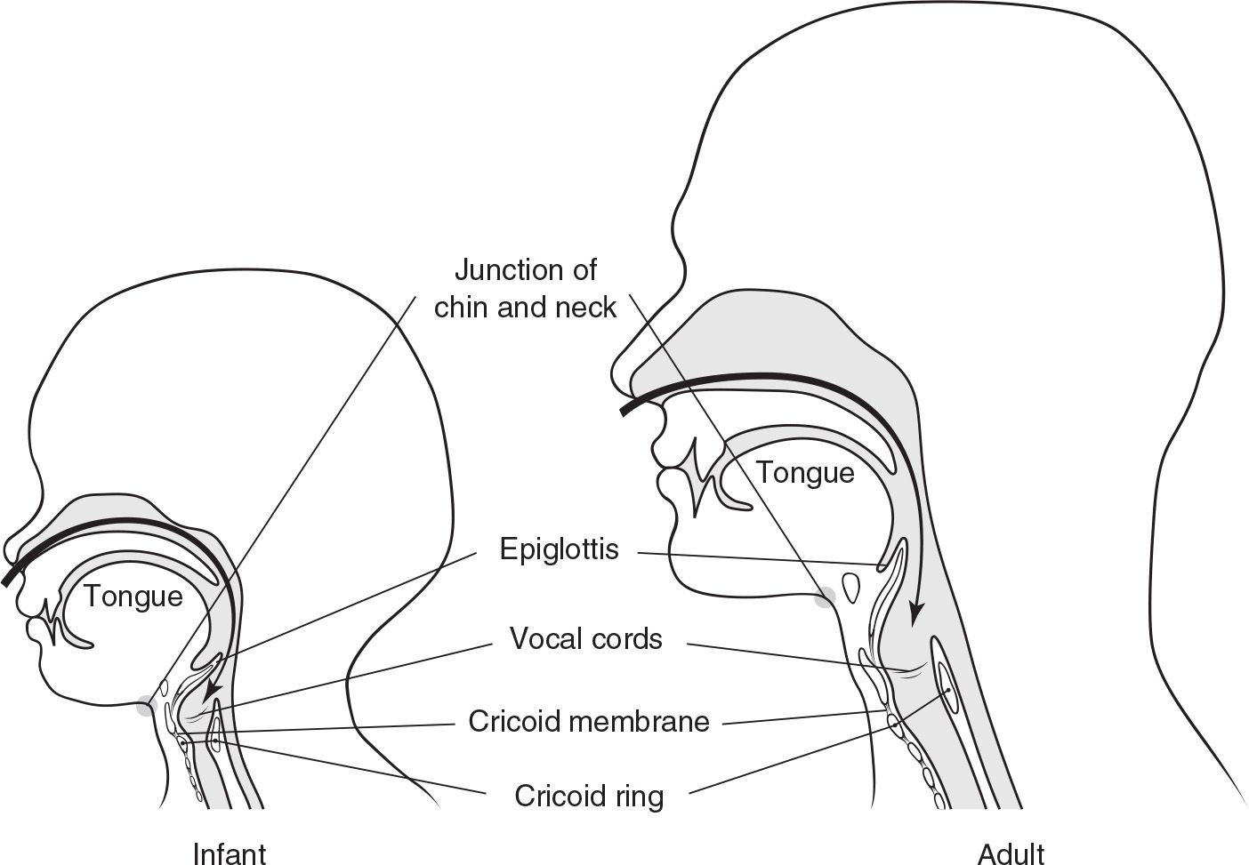
FIGURE 89.1 The anatomic differences particular to children are (a) higher, more anterior position of the glottic opening (note the relationship of the vocal cords to the chin/neck junction); (b) relatively larger tongue in the infant, which lies between the mouth and the glottic opening; (c) relatively larger and more floppy epiglottis in the child; (d) the cricoid ring is the narrowest portion of the pediatric airway versus the vocal cords in the adult; (e) position and size of the cricothyroid membrane in the infant; (f) sharper, more difficult angle for blind nasotracheal intubation; (g) larger relative size of the occiput in the infant.
 General Basic Steps
 Preparation
 Preoxygenation
 Pretreatment
 Protection and positioning
 Paralysis and induction
 Placement of tube and proof of tube placement
 Postintubation management
TECHNIQUE
If crash airway and difficult airway algorithms are not indicated, then rapid sequence intubation (RSI) is the preferred approach. This approach is summarized in seven discrete steps, each beginning with the letter “P.”
 Preparation: Directed history, physical examination, indications/contraindications for RSI
 Assemble equipment using the “SOAP ME” mnemonic (TABLE 89.1)
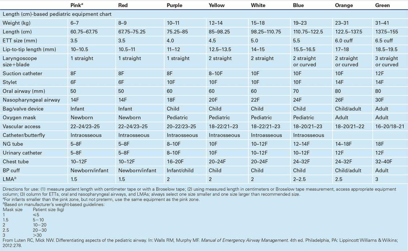
 Size is best estimated using Broselow tape or centimeter measuring tape
 Oral airway
 Size using Broselow tape or distance from the angle of the mouth to the ear tragus
 Nasopharyngeal airway
 Size using Broselow tape, distance from the tip of the nose to the ear tragus, or largest comfortable size that does not produce skin blanching
 Laryngoscope blade
 Straight/Miller blade traditionally has been preferred to the curved blade for infants and young children. However, either blade can be used in any age group depending on availability and operator comfort.
 Endotracheal tube (ETT) size based on Broselow tape or calculated as follows:
 Uncuffed: (Age in years/4) + 4 (subtract 0.5−1 for cuffed tube)
EQUIPMENT FOR RSI—“SOAP ME” MNEMONIC |
S | Suction | Yankaur device (children/adolescents) and/or flexible catheters (infants), suction tubing, wall-mounted suction |
O | Oxygen | Face mask (preferably nonrebreather), oxygen tubing, high-flow oxygen source, Bag/Valve device (with positive-pressure valve) |
A | Airway | Laryngoscope handle with functional light source and blades, endotracheal tubes, airway tape, stylets, oral/nasopharyngeal airways of varying sizes. Rescue equipment (e.g., Bougie, GlideScope, LMA, cricothyrotomy kit, etc.) should be available in case RSI fails. |
P | Pharmacology | Weight-based medications should be prepared in advanced. Agent selection will depend on circumstances and may include sedatives, induction agents, neuromuscular-blocking agents, lidocaine, and atropine. |
ME | Monitoring equipment | Cardiorespiratory monitoring with pulse oximetry and frequent blood pressure checks through postintubation monitoring phase. Following endotracheal tube placement, secondary confirmation with end-tidal CO2 calorimeter (qualitative/semiquantitative) and/or capnography (quantitative) |
LMA, laryngeal mask airway; RSI, rapid sequence intubation. | ||
 Historically, uncuffed tubes were preferred in infants and young children due to high rates of subglottic stenosis. Currently, either tube may be used in any age group if leak pressures are monitored.
 Prepare extra tubes, both 0.5 size smaller and larger than estimated
 A stylet can be used to provide rigidity (TABLE 89.2)
 ETT depth by Broselow tape or calculated (if age >1 year)
 Formula (in cm): (Age in years/2) + 10 or Tube size × 3
 End-tidal CO2 monitor
 If weight <15 kg, use pediatric calorimeter to avoid false negative readings
 Have airway alternatives available (e.g., GlideScope, Airtraq, laryngeal mask airway [LMA], Bougie, needle cricothyrotomy equipment)
 Preoxygenation
 Theoretically, deliver 100% oxygen for 3 minutes. Practically, use nonrebreather facemask (with positive end-expiratory pressure [PEEP] valve) and high-flow nasal cannula once RSI is considered.
 If child becomes apneic, use bag valve mask (BVM) ventilation prior to intubation
 Perform neck extension and E-C clamp technique with bag-mask ventilation (BMV) if C-spine injury is not suspected
 If two providers are available, one person maintains mask seal while the other compresses the bag
 Use the rhythm “squeeze, release, release” to allow time for exhalation
 Insert an oral airway in an unconscious patient who is difficult to ventilate
 Pretreatment: Refers to the administration of medications to attenuate the potential adverse effects of intubation (TABLE 89.3)
 Prior recommendations summarized by “LOAD” (Lidocaine, Opioid, Atropine, Defasciculating agent)
 No pretreatment agent is recommended routinely for pediatric RSI
 Lidocaine: May limit further rise in ICP in cases of head trauma or elevated ICP
 No data to suggest or refute use to prevent reflex bronchospasm
 Fentanyl: Analgesic effects may decrease the reflex sympathetic response
 May cause hypotension or respiratory depression with other sedatives
 Atropine: Used for its anticholinergic effects to prevent or treat bradyarrhythmias
 Antisialogogue effect is delayed, limiting its use in RSI
 Interferes with pupillary response of the neurologic examination after paralysis
 Defasciculating agent: “Defasciculating” and “Priming” doses are no longer recommended
|



Full access? Get Clinical Tree


