 Confirmation of the diagnosis of obstructive uropathy in patients suspected of ureteral colic
 Abdominal or flank pain
 Hematuria
 Groin pain
 Acute urinary retention
 Known or suspected acute renal failure
 Laboratory evidence of renal failure
 Oliguria or anuria
 Painless hematuria or proteinuria
 Suspected renal abscess
 Infected urine with fever and abdominal or flank pain
 Suspected or known abdominal trauma
CONTRAINDICATIONS
 None
None
RISKS/CONSENT ISSUES
 Allergy to the ultrasonography gel
LANDMARKS
 The right kidney is usually located inferior and posterior to the liver
 The left kidney is located inferior to the spleen
 The bladder should be imaged in the suprapubic region
TECHNIQUE
 A 3.5-MHz probe is commonly suitable for most adults, although a 5.0-MHz probe can be used in patients with a thinner body habitus and in children
 The Right Kidney
 With the patient supine, start at the midaxillary line at the level of the lower ribs holding the probe in the longitudinal axis or slightly oblique and scan laterally until the sagittal view of the hepatorenal space (Morison pouch) and the right kidney are visible
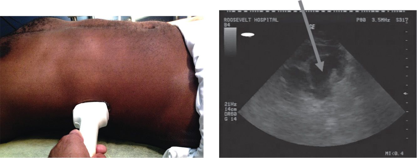
 Rotate the probe 90 degrees to obtain a transverse image of the kidney. Move the probe superiorly and inferiorly to locate the renal hilum and visualize the full extent of the parenchyma (FIGURE 40.1).
 The Left Kidney
 The left kidney is most easily located by placing the probe hand against the bed while scanning the left flank at the posterior axillary line at the level of the lower ribs
 Rotate the probe 90 degrees to obtain a transverse view of the left kidney
 The Bladder
 The bladder is best imaged when it is moderately filled at the time of examination
 Place the probe suprapubically in the transverse plane. Angle the probe toward the patient’s feet. Color Doppler techniques can be used over the trigone area to verify the presence of ureteral flow jets indicating urine flow into the bladder.
 Rotate the probe 90 degrees to obtain a sagittal view



Full access? Get Clinical Tree


