Ultrasound-Guided Proximal Obturator Block: A Proximal Interfascial Technique
Ahmad Mouhammad Taha
 Background: The obturator nerve is a branch of the lumbar plexus. It arises from the anterior divisions of lumbar nerves 2, 3, and 4. The obturator nerve usually divides into anterior and posterior branches before it enters the obturator canal, although occasionally it splits from within the canal. After both obturator branches exit the pelvis, they lie close together for a short disTance, between the pectineus muscle (superiorly) and the obturator externus muscle (inferiorly) (Fig. 26.1), where both branches can be blocked using a single injection. Distally, the two branches diverge. The anterior branch runs lateral, anterolateral, and then anterior to the adductor brevis muscle, whereas the posterior branch runs deep. The obturator nerve supplies the hip joint (via its hip branch), the knee joint (via the posterior branch), the adductor muscles (via the anterior and posterior branches), and a highly variable skin area on the medial aspect of the thigh (via the anterior branch).
The main obturator nerve and its two branches are flat nerves and difficult to identify. Even with combined ultrasound nerve stimulation guidance, multiple needle passes are usually required. On the contrary, the facial border of the adductor muscles can be easily identified. On the ultrasound image, the hyperechoic fascial borders of the pectineus, adductor longus, and brevis muscles usually form a cHaracteristic tricompartmental configuration; this visually resembles the letter Y with its stem directed posteriorly (Fig. 26.2).
 Indications
Knee and above-knee surgeries
Transurethral resection of bladder tumor (TURT)
Chronic hip pain
When prolonged thigh tourniquet inflation is required
 Transducer type: Linear probe (10 to 13 MHz).
 Needle: Short-beveled, 5 cm. A longer needle may be required for obese patients or when an in-plane technique is used.
 Local anesthetics: 10 to 15 mL of 2% lidocaine, 0.5% ropivacaine.
 Patient position: Supine. The thigh is abducted and externally rotated.
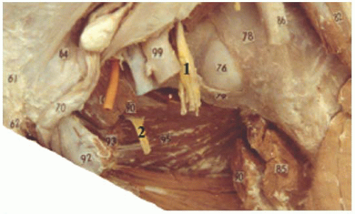 Figure 26.1. After the obturator nerve (2) exits the pelvis, it lies between the pectineus muscle (cut) (90) superiorly and the obturator externus (95)
Get Clinical Tree app for offline access
Related posts: Microanatomy of the Peripheral Nervous System and Stimulation Thresholds Inside and Outside the Epineurium Microanatomy of the Peripheral Nervous System and Stimulation Thresholds Inside and Outside the Epineurium
 Ultrasound-Guided Intraneural Injection—The Human Data Ultrasound-Guided Intraneural Injection—The Human Data
 Ultrasound-Guided Blocks at the Elbow and Forearm Ultrasound-Guided Blocks at the Elbow and Forearm
 Ultrasound-Guided Anterior Sciatic Nerve Block Ultrasound-Guided Anterior Sciatic Nerve Block
 Ultrasound-Guided Caudal, Lumbar, and Epidural Injection Ultrasound-Guided Caudal, Lumbar, and Epidural Injection
 Ultrasound-Guided Genitofemoral Nerve Block Ultrasound-Guided Genitofemoral Nerve Block

Full access? Get Clinical Tree


|