Criteria
Mild
Moderate
Severe
Neuroimaging
Normal
Normal or abnormal
Abnormal
Initial GCS
13–15
9–12
<9
Loss of consciousness
Absent or upto 30 min
30 min to 24 h
More than 24 h
Post-traumatic amnesia
Absent or upto 24 h
1–7 days
More than 7 days
Change in mental status
Absent or upto 24 h
More than 24 h
Prehospital and Emergency Department
Two major causes of secondary brain injury after TBI are cerebral hypoperfusion and hypoxia. Studies have shown that systolic BP < 90 mmHg and PaO2 < 60 mmHg are associated with poor outcomes after TBI [1]. The prehospital treatment is focused toward establishing and maintaining adequate circulation, patent airway and oxygenation. Securement of airway by endotracheal intubation should be considered in patients with GCS <9 while ensuring hemodynamic stability. Crystalloids are preferred over colloids for fluid resuscitation as use of albumin resulted in increased mortality in a randomized clinical trial [2]. All patients with suspected moderate to severe TBI or GCS <15 should undergo neuroimaging evaluation. Non-contrast CT of head is preferred modality due to rapidity, wide availability and high sensitivity for detection of intra- and extraparenchymal hemorrhage and fractures. Common findings on initial CT of head in severe TBI include one or more of cerebral contusion (frontal and temporal lobes are common location), subdural hematoma, epidural hematoma, subarachnoid hemorrhage, intraventricular hemorrhage, diffuse cerebral edema, skull fracture and extracranial hematoma. Frequently, initial CT scan in comatose patients with severe TBI and diffuse axonal injury may be largely unremarkable. A follow up CT done 6–12 h after the initial CT may show development of new lesions or expansion of previously seen contusions. Early detection and treatment of intracranial hypertension should begin in emergency department as both duration and severity of low cerebral perfusion are associated with worse outcomes. Patients with severe TBI should be transferred to a tertiary center with emergent neurosurgical services once hemodynamic stability is established [3] (Fig. 40.1).
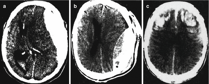
Fig. 40.1
CT scan of the head showing different findings in severe traumatic brain injury. (a) Crescent shaped hyperdensity in the suggestive of left sided acute subdural hematoma with mass effect and left to right midline shift. (b) Lenticular hyperdensity suggestive of acute epidural hematoma with effacement of left lateral ventricle. (c) Bifrontal traumatic cerebral contusions
Mechanical Ventilation
Positive end-expiratory pressure should be kept to minimum to adequately oxygenate in patients with intracranial hypertension requiring mechanical ventilatory support. Prolonged hyperventilation induced hypocapnia increases risk of ischemia by cerebral vasoconstriction while permissive hypercapnia may lead to intracranial hypertension via cerebral vasodilation.
Analgesia, Sedation and Neuromuscular Paralysis
Inadequate pain control, agitation and anxiety may increase ICP and cerebral oxygen demand while unnecessary and excessive sedation results in inability to detect change in neurological exam. Therefore, judicious use of short acting opioids such as fentanyl, ramifentanyl or morphine is warranted for pain control and propofol or short acting benzodiazepine such as midazolam may be used to provide sedation. Non-depolarizing muscle relaxants should be used for patient-ventilator dyssynchrony causing refractory hypoxia and to treat intracranial hypertension caused by excessive coughing or straining.
Surgical Treatment
Epidural Hematoma
Patients with EDH and GCS score <9 should undergo surgical evacuation. Surgery is also recommended for patients with EDH volume ≥30 cc, thickness ≥15 mm or midline shift of ≥5 mm regardless of GCS. Patients managed nonoperatively should undergo serial CT imaging and frequent neurological monitoring where emergent neurosurgical treatment is available. Temporal location increases the risk of deterioration and therefore, threshold for surgery should be kept lower for patients with temporal EDH. Time from neurological worsening to EDH evacuation correlates more with outcomes than time from injury to evacuation. Therefore, in patients undergoing initial non-surgical treatment, neurological deterioration as defined by worsening level of consciousness, abnormal papillary reflex, and appearance of new focality or worsening of existing focal deficits should prompt urgent surgical evacuation.
Subdural Hematoma
The indications for surgery in acute traumatic SDH include clot thickness >10 mm or midline shift >5 mm on initial CT imaging regardless of GCS. In patients with smaller SDH, GCS <9 or decrease in GCS by 2 points or more since presentation, presence of papillary abnormality or persistent elevation of ICP >20 mmHg should prompt surgical evacuation.
Intraparenchymal Hematoma/Traumatic Cerebral Contusion
Patients with GCS of <9 with frontal or temporal hematoma of >20 cc with midline shift of ≥5 mm or cisternal compression and those with any hematoma of >50 cc should undergo evacuation. Also, patients with neurological deterioration thought to be related to intraparenchymal hematoma should also be treated surgically [4]. Neurologically stable patients with parenchymal contusion showing no significant mass effect on CT scan can be managed nonoperatively with close monitoring and serial imaging. Patients with medically refractory intracranial hypertension from diffuse cerebral edema may be treated with bifrontal or hemispheric decompressivecraniectomy.
Evidence Contour
Indication of Intracranial Pressure Monitoring and Pressure Threshold
Invasive ICP monitoring is not routinely indicated in all cases of TBI and the risks of monitoring such as infection and bleeding must be weighed against the benefit of additional information obtained by such monitoring. Patients with severe TBI with abnormal CT scan and GCS <9 should undergo ICP monitoring as they have high likelihood of transient or persistent elevation in ICP resulting in compromise in cerebral perfusion pressure and inability of serial clinical examinations to identify subtle changes related to intracranial hypertension due to poor baseline neurological status. Normal neuroimaging especially during early hours of TBI does not rule out intracranial hypertension. Patients with two or more of the following factors should undergo ICP monitoring in the absence of abnormal findings on CT scan of the head: age above 40 years, motor posturing, systolic blood pressure <90 mmHg. The ICP threshold above which interventions aimed at lowering the ICP should be implemented is unclear at this time. ICP of above 20 mmHg is generally recommended as the treatment threshold by several guidelines. Cerebral perfusion pressure (CPP = MAP-ICP) based threshold may provide for a more physiologically rational parameter and should be used in conjunction with ICP parameter with goal CPP >60–65 mmHg. Different devices used for ICP monitoring use any of available technologies such as fiberoptic sensor, microchip with internal strain gauge, air pouch or fluid filled catheter connected to pressure transducer. Devices also differ in terms of location of tip such as subdural, epidural, subarachnoid, intraparenchymal and intraventricular. Of these, fluid-filled transduced ventriculostomy catheter provides the most accurate ICP value and also allows for therapeutic CSF drainage and is therefore, preferred over other modalities.
Related posts:


Full access? Get Clinical Tree


