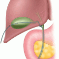
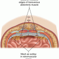
Fig. 21.1
Normal superficial venous anatomy
Vein surgery is appropriate for patients manifesting classic venous symptoms of aching leg pain with edema, visible varicose veins, and duplex ultrasound proven superficial venous incompetence.
Preoperative Preparation
A presurgical trial of elastic compression with symptom improvement is predictive of successful surgical results. All patients should have duplex sonographic proof of reflux greater than 0.5 s and saphenous vein diameter >5 mm. Immediately prior to surgery mapping the varicosities with the patient standing facilitates phlebectomy.
Operative Strategy
There are two components to surgery, which may be staged, by surgeons preference
1.
Elimination of reflux: endovenous ablation vs. open stripping
2.
Phlebectomy of varicosities
Operative Technique (Fig. 21.2)

Fig. 21.2
Endovenous catheter mode of action
1.
Endovenous ablation using radiofrequency vs. laser energy. Although the hardware required differs somewhat the procedure is similar and entails:
Ultrasound mapping of the GSV—facilitated by reverse Trendelenburg position.
Accessing the GSV at the knee and insertion of a sheath to facilitate device insertion. The vein is accessed under ultrasound control using a micro catheter cannulation sheath and then a working sheath of 7F (RFA) or 5F (Laser) is inserted using Seldinger technique. The device is inserted no closer than 2 cm from SaphenoFemoral Junction (SFJ).
Tumescent anesthesia is infiltrated under ultrasound control by hand or using an infusion pump.
The patient is positioned in Trendelenburg and the device is activated per manufacturer’s protocol.
Completion ultrasonography to confirm successful treatment and to rule out deep venous thrombosis.
Full access? Get Clinical Tree


