 Trephination: To create a fistula through the nail to the hematoma
 Decompression, drainage, and pain relief of small subungual hematomas
 Nail removal and nail bed laceration repair
 Large hematomas and nail bed lacerations
 Partial nail avulsion or subluxation with nail instability or nail fold disruption
CONTRAINDICATIONS
 Significant crush injuries or missing/destroyed nail matrix warrant specialty consultation
RISKS/CONSENT ISSUES
 Germinal matrix injuries and open tuft fractures should be documented and referred to a specialist to ensure optimal outcome
LANDMARKS
 Hematomas typically collect on the sterile matrix under the nail
 Germinal matrix
 Region where new nail is formed
 Avoid injury during the procedure
 General Basic Steps
 X-ray digit if fracture possible
 Prepare patient
 Consider anesthesia
 Perform procedure (FIGURE 73.1)
 Trephination
 Goal is to form a hole through the nail of sufficient size to drain the hematoma
 Personal protection (including an eye shield) as blood may spurt out when released
 Needle or scalpel method: Apply gentle pressure with the tip of the instrument perpendicular to the surface of the nail, twisting until blood is released
 Heated paper clip method: Creates a wider hole but may tattoo the nail bed
 Disposable electrocautery device method: Quick and effective (FIGURE 73.2)
 Discharge instructions are to soak the finger in warm water twice a day for 7 days to allow the blood to continue to drain
 Nail Bed Laceration Repair
 Supplies: Iris scissors, hemostats, and suture set with fine absorbable sutures (5-0 to 7-0 chromic or Vicryl)
 Perform digital block under sterile conditions
 Apply tourniquet for hemostasis
 Remove the nail from the nail bed matrix
 Insert closed Iris scissors horizontally under the nail
 Gently spread scissors and advance in repeated movements, gradually progressing to the nail root and separating the entire nail from the nail bed
 Once the nail is free from the nail bed and eponychium, grasp with hemostat and gently pull longitudinally to free the nail
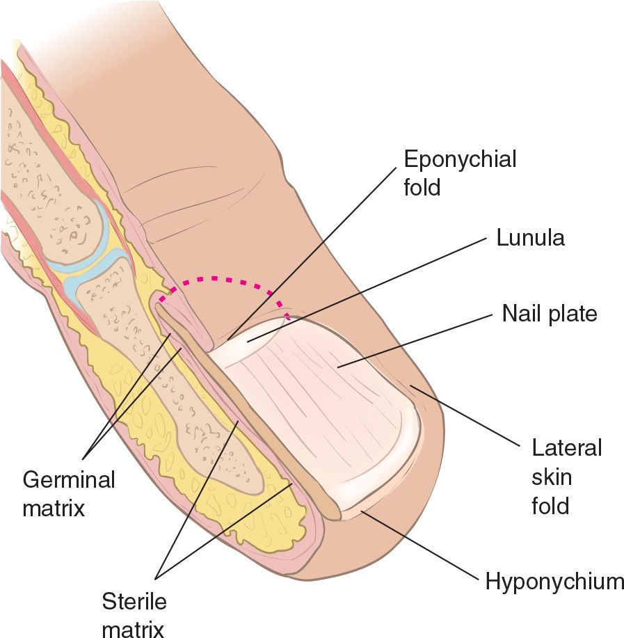
FIGURE 73.1 Anatomy of the finger and nail bed. (From Eberlein R. Hand and finger injuries. In: Henretig FM, King C, eds. Textbook of Pediatric Emergency Procedures. Philadelphia, PA: Williams & Wilkins; 1997:1048, with permission.)



Full access? Get Clinical Tree


