Key Clinical Questions
Introduction
Cellulitis is an acute infection involving the epidermis, dermis, and subcutaneous tissues. Most cases are caused by Staphylococcus aureus or beta-hemolytic streptococci, although in many patients with presumed infectious cellulitis, a definite bacteriologic diagnosis cannot be made. Occasionally, cellulitis is due to nonbacterial pathogens or noninfectious inflammatory conditions. The costs to society of cellulitis are significant. Up to 2% of hospital admissions are due to cellulitis. While only 7% of patients with cellulitis are admitted to the hospital, hospitalized patients account for 80% of the total health care costs associated with cellulitis.
Pathophysiology
Healthy skin is resistant to infection. Skin breakdown is usually required for cellulitis to develop. This may be obvious or too subtle to detect. Tinea pedis is an especially common portal of entry for bacteria in lower-extremity cellulitis. Other local risk factors for cellulitis include common skin conditions such as eczema and psoriasis, trauma, intravenous drug use, animal and human bites, venous stasis, and lymphedema. Medical risk factors for cellulitis include diabetes, arterial insufficiency, cirrhosis, renal insufficiency, and neutropenia.
Cellulitis occasionally causes bacteremia. Rarely, cellulitis arises from systemic bacteremia, with secondary seeding of the skin. This is more common in neutropenic patients. These infections lead to bacterial proliferation in the vessel wall, tissue ischemia, and skin necrosis. Clinically, this presents as an area of inflamed skin, with a hemorrhagic pustule that develops into a necrotic ulcer (ecthyma gangrenosum).
Clinical Presentation
Cellulitis presents with pain, swelling, and erythema of the skin. In most patients with cellulitis, the margins of erythema are not sharply demarcated. Fever is usually present in patients admitted to the hospital with cellulitis, although it is often absent in milder forms. A minority of patients with cellulitis have enlargement of the draining lymph nodes (lymphadenitis). The bacteriology of cellulitis usually cannot be inferred from the physical examination. However, suppuration and abscess formation is strongly suggestive of Staphylococcus aureus. Rapid clinical evolution, lymphangitis, and features of erysipelas (discussed later), such as peau d’orange and a raised, well-demarcated border, suggest beta-hemolytic streptococci. A number of epidemiologic features in cellulitis put patients at risk for particular pathogens. These are summarized in Table 203-1 and discussed in more detail later in this chapter. As well, several noninfectious conditions may mimic cellulitis. These are summarized in Table 203-2.
| Exposure | Pathogen |
|---|---|
| Dog bite | Pasteurella multocida, Capnocytophaga sp., mixed aerobic and anaerobic flora |
| Cat bite | Pasteurella multocida, mixed aerobic and anaerobic flora |
| Fish exposure | Mycobacterium marinum, Erysipelothrix rhusiopathiae, Streptococcus iniae |
| Salt water | Vibrio vulnificus |
| Fresh water | Aeromonas hydrophilia, Edwardsiella tarda, Chromobacterium violaceum, protothecosis |
| Cirrhosis | Vibrio vulnificus from raw seafood consumption; gram-negative rods |
| Intravenous drug use | Eikenella corrodens and other oral flora, Pseudomonas aeruginosa and other gram-negative rods, anaerobes including Clostridium sp. |
| Neutropenia | Pseudomonas aeruginosa and other gram-negative rods fungi |
| Condition | Distinguishing Features |
|---|---|
| Venous stasis | Erythematous, swollen, warm lower extremity with dermatitis and hyperpigmentation; typically bilateral in obese individuals, and unilateral when occurring after saphenous vein harvest or deep venous thrombosis; skin breakdown and ulceration may develop at the medial and lateral malleoli; if there is no bacterial superinfection, fever and leukocytosis will be absent, and clinical improvement occurs with limb elevation and other measures to reduce edema. |
| Arterial insufficiency | Nonhealing foot wounds with dependent rubor may mimic cellulitis; patients may also have claudication, rest pain, and diminished or absent pedal pulses; if suspected, noninvasive vascular studies, such as ankle-brachial index, should be performed. |
| Superficial thrombophlebitis | Swelling, induration, and erythema, typically at an intravenous site; palpable cord usually present; fever and leukocytosis absent if no superinfection. |
| Deep venous thrombosis | Unilateral swollen leg; low-grade fever may be present; superficial veins may be congested, and a cord may be palpable, but physical examination not sensitive; if suspected, obtain duplex ultrasonography. |
| Contact dermatitis | Weeping skin with vesicles and erythema; well-localized lesion, sometimes with pattern suggestive of irritant exposure; superinfection may occur. |
| Herpetic whitlow | Swollen painful finger with vesicles; patients are often health care workers, such as dental hygienists or intensive care nurses, who come into contact with the oral cavity and saliva of patients. |
| Pyoderma gangrenosum | Ragged undermined ulcer, typically on the lower extremity; cultures usually positive as heavily colonized with bacteria; common in inflammatory bowel disease; obtain dermatology consult and biopsy if suspected. |
| Insect bite | Rapid and sometimes impressive local swelling and erythema, without fever or systemic toxicity; good response to antihistamines; rarely, with spider bites, central necrosis develops. |
| Erythema nodosum | Hot, warm, tender plaques (panniculitis) involving the shins; seen with sarcoid, rheumatologic disease, fungal infection. |
| Sweet syndrome | Papules, plaques, and panniculitis involving the head, neck, upper trunk and arms; fever and neutrophilia often present; may be postinfectious syndrome in young person, or a harbinger of hematopoietic malignancy in an older one. |
| Gout | Exquisitely tender arthritis, often with prominent soft tissue inflammation, involving the great toe, midfoot, knee, and other joints; fever and leukocytosis often prominent; tophi may be present; may complicate hospital admission for other illness. |
| Calciphylaxis | Painful plaques on the extremities, which may become necrotic; typically occurs in hemodialysis patients with extensive atherosclerosis and high calcium-phosphate product. |
| Familial Mediterranean fever | Recurrent, brief, self-limited attacks of fever, serositis and erysipelas-like skin erythema; patients are typically of Mediterranean or Middle Eastern descent, and have a positive family history. |
Bacterial cellulitis should respond to antibiotic therapy after several days of treatment. Disease progression or failure to improve usually reflects infected fluid collections or involvement of tissue deep to the skin. Involvement of deeper tissues and structures should also be suspected in cases of systemic toxicity or disproportionate pain (necrotizing fasciitis), foot ulcers in diabetics (osteomyelitis), cellulitis overlying a joint replacement (infected prosthetic joint), or cellulitis of the abdominal wall (bowel inflammation or enterocutaneous fistula) (Table 203-3).
|
The yield of blood cultures is low in healthy patients with cellulitis, but these should be performed in patients with high fever, systemic toxicity and sepsis, or compromised immunity. Wounds, pustules, and abscesses should be cultured, if present. These have a diagnostic yield of around 10% to 20%. Complete blood count, electrolytes, blood urea nitrogen, creatinine, and glucose should be performed in sicker patients or those with comorbid conditions. An elevated creatine kinase may heighten the clinical suspicion of necrotizing fasciitis or clostridial myonecrosis. Imaging studies have a limited role in the management of cellulitis. Plain radiographs may reveal a foreign body or gas in the tissues; computed tomography (CT) and magnetic resonance imaging (MRI) may assist in the diagnosis of necrotizing fasciitis.
There are important variants of cellulitis with either distinctive clinical features, bacteriology, or both. These are discussed below.
Erysipelas and recurrent cellulitis in the absence of a wound are both generally due to streptococci. In erysipelas (Figure 203-1), there is skin edema with dimpling similar to the appearance of an orange peel (peau d’orange). Inflammation involving lymphatic drainage may be present as erythematous lines directed to regional lymph nodes (lymphangitic streaking). The draining nodes may be enlarged and tender. Abscesses and chronic skin ulcers are absent. The most typical laboratory abnormality is a polymorphonuclear leukocytosis. The microbiology is usually not determined; rarely blood cultures are positive. The distinctive appearance, occasional positive blood culture, molecular diagnostic studies, and the observation that penicillin is effective therapy lead to the conclusion that these infections are caused by beta-hemolytic streptococci. Group A and group G streptococci are responsible for most cases, group C and group B streptococci account for the remainder.
Figure 203-1
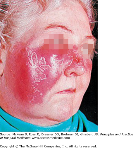
Erysipelas with typical clinical features (superficial, fiery-red, well-demarcated, indurated dermal cellulitis with peau d’orange). Most cases involve the cheeks or extremities. Beta-hemolytic streptococci are responsible. (Reproduced, with permission, from Fauci AS, Braunwald E, Kasper DL, et al. Harrison’s Principles of Internal Medicine. 17th ed. New York: McGraw-Hill; 2008, Fig. 130-4.)
Recurrent cellulitis is often related to underlying anatomic abnormalities. The area of involvement in any given patient is consistent from episode to episode. Many patients have recurrent leg cellulitis in the setting of edema from chronic venous or lymphatic insufficiency, with morbid obesity an increasingly common underlying problem. In these patients, inflammatory changes from chronic venous stasis need to be distinguished from acute cellulitis. Other common sites are the chest wall or the ipsilateral arm after mastectomy, or in the lower extremity after saphenous vein removal for vascular grafting. Onset is sudden, with local pain and rapidly progressive erythema. Associated symptoms include fever, headache, nausea, and myalgias. The time from onset to fully developed cellulitis can be as short as a few hours. Diabetics with arterial insufficiency may also have recurrent cellulitis, often from a persistent focus of infection, such as osteomyelitis, as discussed below.
Necrotizing fasciitis is a rapidly progressive infection centered about the deep fascial tissues. Patients complain of severe pain at the site of involvement and frequently present with sepsis syndrome. Early in the course, the overlying skin may appear normal. Later, there is erythema with a waxy unhealthy congested appearance. In the final stages, after thrombosis of the underlying subcutaneous vessels, there is violaceous discoloration and blistering. The severe pain and ill appearance of these patients differentiate them from those with more superficial infection. In unstable patients, surgical exploration with immediate histopathologic review of a biopsy sample is standard care. When the diagnosis is uncertain and the patient is well enough to be transported, imaging may be helpful. CT is more sensitive for gas in the soft tissues (this finding is often absent in necrotizing fasciitis), and MRI is more sensitive for fascial edema. However, both imaging modalities are imprecise in this setting, and the decision to proceed with surgical exploration should be based primarily on clinical grounds.
The soft tissues around the eye are separated from the orbit by the septum, a tough membrane of protective connective tissue. Preseptal, or periorbital, cellulitis is much more common than postseptal, or orbital, cellulitis. Preseptal cellulitis arises from local skin breakdown and inoculation, similarly to cellulitis elsewhere. Postseptal cellulitis may arise from sinusitis, eye surgery, trauma, periodontal infection, and occasionally preseptal cellulitis. Preseptal and postseptal cellulitis may be difficult to distinguish at the bedside. Orbital cellulitis may be diagnosed if proptosis, diplopia, vision loss, and limitation of eye movement are present. The bacteriology of preseptal cellulitis is generally similar to that of other sites. Haemophilus influenzae type b was once a common cause of periorbital cellulitis in toddlers, but has become rare with universal childhood vaccination. Orbital cellulitis may involve gram-negative rods and anaerobes, as well as staphylococci and streptococci, especially if it arises from a sinus or odontogenic source. In neutropenic patients or those with diabetic ketoacidosis, mucormycosis should be suspected, and urgent otolaryngology consult should be obtained. CT scan should generally be obtained to delineate the extent of orbital involvement. Ophthalmology consultation is advised if orbital involvement is suspected.
Related posts:
 Strategies for Cost-Effective Care
 Building, Growing, and Managing a Hospitalist Practice
 Designing a Hospitalist Compensation and Bonus Plan
The Face of Health Care Emerging Issues for Hospitalists
Designing a Hospitalist Compensation and Bonus Plan
The Face of Health Care Emerging Issues for Hospitalists
 Medical Malpractice
Preventing and Managing Adverse Patient Events: Patient Safety and the Hospitalist
Medical Malpractice
Preventing and Managing Adverse Patient Events: Patient Safety and the Hospitalist

Full access? Get Clinical Tree


