 To evacuate abnormal collections of fluid from the joint space for synovial fluid analysis, especially in the investigation of the following conditions:
 Septic arthritis
 Crystal arthropathy
 Hemarthrosis
 Inflammatory processes
 To diagnose occult fracture or ligamentous injury
 To decrease/relieve pressure in the joint for pain relief
 To assess whether an overlying laceration extends into the joint space using methylene blue
 To instill medication for treatment and pain relief
CONTRAINDICATIONS
 Absolute Contraindications
 Abscess/cellulitis in the tissue overlying the puncture site
 Relative Contraindications
 Known bacteremia
 Bleeding diathesis or anticoagulant therapy
 Prosthetic joint
 General Basic Steps
 Patient preparation
 Sterile technique
 Analgesia
 Aspiration
LANDMARKS
 Anterior Approach
 Coracoid process and head of the humerus
 Posterior Approach
 Posterolateral edge of the acromion
SUPPLIES
 Povidone–iodine or other antiseptic solution, drapes
 Lidocaine with epinephrine
 25- and 18-gauge needles, 20-mL syringe
 Gauze pads, adhesive tape
TECHNIQUE
 Patient Preparation
 The patient should sit upright with arm in slight external rotation
 Confirm landmarks and, if needed, mark the needle insertion point
 Sterilize the needle insertion area with povidone–iodine solution or comparable skin antiseptic
 Wipe injection site with alcohol to avoid introduction of iodine into synovium
 Drape the area with sterile towels
 Analgesia
 Use a 25-gauge needle to raise a wheal of anesthetic
 Inject lidocaine with epinephrine at the puncture site
 Anesthetize the subcutaneous tissue and a track toward the joint
 Avoid entering the joint space at this point if synovial fluid analysis is desired
 Aspiration
 Anterior Approach
 Insert an 18-gauge needle attached to a 20-mL syringe just below and lateral to the coracoid process, medial to the head of the humerus
 Point the needle posterolaterally to avoid the joint capsule
 Gently aspirate while advancing the needle. The needle should be advanced approximately 3 cm or until fluid is aspirated (FIGURE 58.1).
 Posterior Approach
 Insert an 18-gauge needle attached to a 20-mL syringe 1 cm below and 1 cm medial to the posterolateral edge of the acromion
 Aim the needle anteriorly toward the coracoid process
 Gently aspirate while advancing the needle. The needle should be advanced approximately 3 cm or until fluid is aspirated (FIGURE 58.2).
 Once the required amount of fluid is obtained, withdraw the needle
 Apply pressure over the area of insertion for 30 seconds or until bleeding stops
 Wipe off all excess povidone–iodine on the skin
 Apply clean dressing
COMPLICATIONS
 Iatrogenic infection
 Excessive pain during procedure
 Localized bleeding
 Reaccumulation of fluid
 Injury to articular cartilage
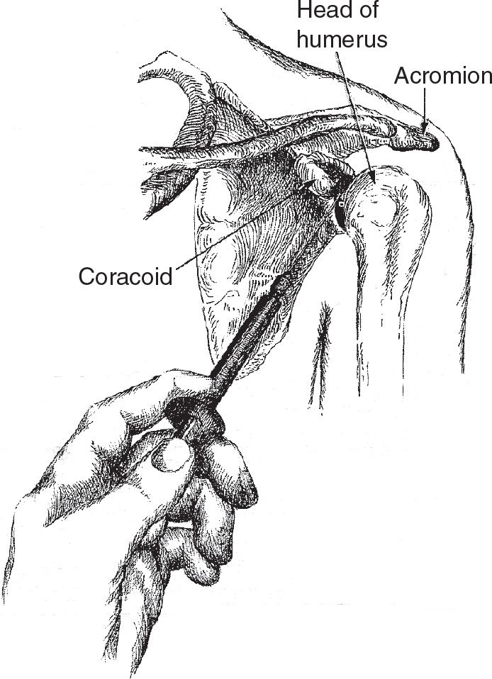
FIGURE 58.1 Anterior approach. (From Simon RR, Brenner BE. Emergency Procedures and Techniques. 4th ed. Philadelphia, PA: Lippincott Williams & Wilkins; 2002:242, with permission.)



Full access? Get Clinical Tree


