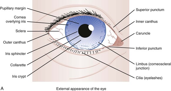
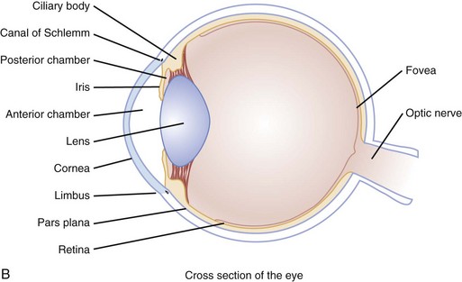
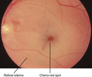
Red and Painful Eye

Chapter 22


Red and Painful Eye


Perspective


Epidemiology and Pathophysiology


Most eye complaints are not immediately sight-threatening and can be managed by an emergency physician. Nontraumatic diseases, such as glaucoma and peripheral vascular disease leading to retinal ischemia, are more common with advancing age. Ocular injuries are the leading cause of visual impairment and blindness in the United States.1 More patients with postoperative complications can be expected to visit the emergency department (ED) as more vision correction surgeries are performed.


The external and internal anatomy of the eye is depicted in Figure 22-1A and B. The globe has a complex layer of blood vessels in the conjunctiva, sclera, and retina. Redness reflects vascular dilation and may occur with processes that produce inflammation of the eye or surrounding tissues. Eye pain may originate from the cornea, conjunctiva, iris, or vasculature. Each is sensitive to processes causing irritation or inflammation.


Diagnostic Approach


Rapid and accurate triage is the most critical consideration in the approach to the red and painful eye. The first question should be, “Did anything get in your eye?” If so, the second question should be, “What do you think it was?” This helps separate trauma from nontrauma, but, more important, seeks to identify quickly eyes that may have been exposed to a caustic substance. Patients exposed to caustic substances require rapid decontamination to prevent permanent loss of visual acuity.


Pivotal Findings


Measurement of the patient’s best corrected visual acuity (i.e., with glasses on, if available) with each eye individually and with both eyes together provides vital information in evaluation of eye complaints. Only a few situations preclude early and accurate visual acuity testing. Eyes exposed to caustic materials should be irrigated as soon as possible. Patients with sudden and complete visual loss in one eye require prompt funduscopic examination to determine the possibility of acute central retinal artery occlusion. This condition is readily apparent as a diffusely pale retina with indistinct or unseen retinal arteries (Fig. 22-2).


Other pivotal findings, which are more likely to be associated with a serious diagnosis, in patients with a red or painful eye are listed in Box 22-1.


History


Chief complaints of pain can be manifestations of a variety of sensations. When carefully questioned, some patients may differentiate among itching, burning, dull pain, sharp pain, and perception of a foreign body. Itching tends to be more often caused by blepharitis, conjunctivitis, or dry eye syndrome. Burning is associated with these conditions and with other mostly superficial problems, such as irritation of a pterygium or pinguecula, episcleritis, or limbic keratoconjunctivitis. A foreign body sensation is more typical of corneal irritation (abrasion, ulcer) or inflammation (keratitis). Sharp pain generally results from abnormalities of the anterior eye, such as keratitis, uveitis, and acute angle–closure glaucoma. Dull pain may be a manifestation of increased intraocular pressure (IOP) or referred from an extraorbital process, such as sinusitis, migraine headache, or temporal arteritis.


A chief complaint of redness commonly results from palpebral or limbal injection of the conjunctiva. However, free blood can be noted behind the bulbar conjunctiva (i.e., subconjunctival hemorrhage) or in the anterior chamber (i.e., hyphema). Both of these can be spontaneous or post-traumatic. Spontaneous subconjunctival hemorrhages may follow coughing or straining. Spontaneous subconjunctival hemorrhage is painless, and the presence of pain raises concern for a more serious cause of the hemorrhage, such as direct globe injury. Often, it occurs without any identifiable precipitating incident and is simply noticed by the patient when looking in a mirror. Hyphema of sufficient size to be noted by the patient or bystander usually arises with pain and blurred vision.


Other subjective findings may be transient and detected only by history. The patient may relate lid swelling, tearing, discharge, crusting, or sensitivity to light. Lid swelling can be caused by inflammatory and noninflammatory processes. Concurrent erythema of the lid favors the former. In the absence of trauma or other external irritant (e.g., contact dermatitis), inflammatory processes include primary lid problems, such as hordeolum (i.e., stye) or blepharitis as well as extension from concomitant conjunctivitis or cellulitis in orbital or periorbital structures. When pain is present, tearing is usually secondary. Discharge and crusting are most commonly associated with conjunctivitis, whether allergic, viral, or bacterial. Blepharitis, dacryocystitis, and canaliculitis are other inflammatory processes that may create a discharge and subsequent crusting.


Other eye status review questions include the following:


• Are contact lenses used? If so, what type, how are they cleaned, and how old are the lenses? Has there been a change in the pattern of use (especially increased use)? Were the lenses worn for a particularly long period recently? Are there problems with the lenses drying out? Does insertion of the lenses worsen or relieve the symptoms? Contact lenses alter the physiology of the cornea, making it relatively hypoxic, drier, and more susceptible to bacterial infection with subsequent ulceration.


• Are glasses worn? If so, when was the last assessment for adequate refraction? When the patient is examined for objective abnormalities in visual acuity, the patient’s subjective interpretation of changes from his or her baseline may be all that is available when corrective lenses are not available or their prescription is out of date.


• Has previous injury or eye surgery occurred? Abnormal examination findings, such as an eccentric or irregularly shaped pupil, may be the patient’s baseline. Pain and redness are expected shortly after eye surgery, but many surgical complications also manifest with a red and painful eye.


• What is the patient’s usual state of health? Several systemic diseases may cause symptoms and signs in and around the eye. Giant-cell arteritis is a vasculitis with subacute systemic manifestations but often acute eye complaints.


• What medications are being taken? Medications that affect the sympathetic or parasympathetic nervous systems may affect ocular physiology, such as aqueous production, or pupil size and reactivity. Bleeding may be potentiated by anticoagulant or antiplatelet medications.


• Are there any known or suspected allergies? Environmental allergens are a common cause of conjunctivitis. Other superficial manifestations can be from chemicals (e.g., contact lens-cleaning solution) or other materials (e.g., makeup).


Physical Examination


A complete eye examination usually includes eight components, although many patients require only a limited or directed eye examination, depending on the presentation. The mnemonic VVEEPP (pronounced “veep”) plus slit-lamp and funduscopic examinations represent these components (Box 22-2). Slit-lamp examination is recommended for any complaint involving trauma and for any medical presentation involving foreign body sensation or alteration of vision. Funduscopic examination is usually pursued if there is visual loss, visual alteration, or suggestion of serious pathology in the history and initial physical examination. A thorough physical examination can be conducted in the following order.


Visual Acuity


The initial determination of a patient’s visual acuity provides a baseline from which deterioration or improvement may be followed. It is also predictive of functional outcome after ocular trauma. Visual acuity is quantitatively assessed by use of a Snellen chart test at a distance of 20 feet (6 m) or a Rosenbaum chart at a distance of 14 inches. Young patients who cannot yet read letters and numbers should be tested with an Allen chart that depicts easily recognizable shapes. Each eye is tested separately, with the opposite eye carefully covered. Patients who do not have their prescribed corrective lenses may be evaluated by having them view the chart through a pinhole eye cover, which negates most refractive errors in vision.


If the patient cannot distinguish letters or shapes on a chart, visual acuity is determined qualitatively. Any printed material suffices. The result may be recorded as, for example, “patient able to read newsprint at 3 feet.” If this is not possible, visual acuity is recorded as:


External Examination


Gross abnormalities are assessed by a visual inspection of both eyes simultaneously. Findings may be more apparent if compared with the opposite side. Fractures of facial bones are associated with ocular injuries, some of which require immediate intervention by an ophthalmologist.2


Globe position is part of the external examination. Subtle exophthalmos and enophthalmos are rare and are best detected by looking inferiorly, tangentially across the forehead, from over the patient’s scalp. Exophthalmos (proptosis) may have traumatic or nontraumatic causes but is a result of increased pressure or a space-occupying lesion within the orbit, which may manifest as pain. Medical causes include cellulitis or intraorbital or lacrimal tumors. Hyperthyroidism may cause enlargement of extraocular muscles. The most important cause of exophthalmos in the ED is retrobulbar hematoma, a condition characterized by hemorrhage within the bony orbit, behind the globe. Orbital compartment syndrome pushes the globe forward, stretching the optic nerve and retinal artery and increasing IOP. The resulting microvascular ischemia is sight-threatening if sufficiently severe and persistent. Orbital emphysema and inflammation caused by a retained foreign body behind the eye are other causes of exophthalmos. The discovery of exophthalmos should prompt ocular tonometry measurements to determine the urgency of intervention. Trauma, particularly penetrating globe injury with extrusion of vitreous, can cause the globe to recede into the orbit, but the most common cause of enophthalmos is actually pseudoenophthalmos when the contralateral globe is proptotic.


Inspection also involves examination of the upper and lower palpebral sulci for foreign bodies or other abnormalities. The lower sulcus is easily viewed after manual retraction of the lower lid toward the cheek and having the patient gaze upward. The upper sulcus is inspected by pulling its lashes directly forward and looking under the lid with white light. The lid can then be everted by pressing a cotton-tipped applicator in the external lid crease and folding the lid margin over the applicator.


Conjunctivitis is a common diagnosis after evaluation of patients with red and painful eyes, but determination of cause is much more difficult on clinical grounds. Patients may be unaware of exposures that could cause a chemical conjunctivitis. Without a known history of environmental allergies or other symptoms and signs to suggest an allergic reaction, allergic conjunctivitis may be mistaken for an infection. However, these are usually managed with removal of any known offending agent and symptomatic relief only. On the other hand, acute infectious conjunctivitis can be bacterial or viral, and management should ideally be directed toward the specific microbiologic cause. The acute management of infectious conjunctivitis accounts for over one-half billion dollars in indirect lost productivity and direct costs of antibiotic prescriptions,3 when many patients do not need them.4


The presence of punctate “follicles” (i.e., hypertrophy of lymphoid tissue in Bruch’s glands) along the conjunctival surfaces of one or both lower lids has been touted to be relatively specific for a nonbacterial (i.e., viral or toxic) cause—though one notable exception is trachoma, a chronic keratoconjunctivitis caused by Chlamydia trachomatis. Indeed, the “typical” viral “pinkeye” has been called acute follicular conjunctivitis.5 Some authorities also believe that the mucoid discharge associated with viral conjunctivitis can be clinically differentiated from the purulent discharge associated with bacterial infection.6 However, most research studying the association of symptoms and signs with positive bacterial cultures have grouped “mucoid or purulent” discharges into one finding, not requiring primary care physician participants to discriminate between the two during data collection.7,8 Thus no experimental evidence supports or refutes these expert opinions.9


Eyelids sticking together, particularly in the morning, is commonly cited as clinical evidence of bacterial, as opposed to viral, conjunctivitis, but this is unreliable. One multicenter primary care study in the Netherlands used logistic regression analysis to conclude that its presence plus the absence of itch and a prior history of conjunctivitis was associated with bacterial conjunctivitis; however, the 95% confidence intervals for the area under the receiver operating characteristic curve ranged from 0.63 to 0.80, thus making it a poor to only fair clinical prediction rule.7 In a single U.S. pediatric ED spanning ages from 1 month to 18 years, a similar analysis found sticky eyelids and either mucoid or purulent discharge to have a positive likelihood ratio of 3.1, and a post-test probability of 96% for positive bacterial cultures in their population; however, this likelihood ratio also indicates only a poor-to-fair test.8 Viral cultures were not performed in either of these studies, so the possibilities of copathogens or bacterial culture of nonpathogenic flora was not assessed. Therefore no good evidence exists for differentiating bacterial from viral causes on clinical grounds.


Pupillary Evaluation


The pupils are inspected for abnormalities of shape, size, and reactivity. These examinations are conducted with light specifically directed into the pupil and by means of the swinging flashlight test.


Blunt or penetrating trauma, previous surgery (e.g., iridotomy for cataract extraction), and synechiae from prior iritis or another inflammatory condition are the most common causes of irregularly shaped pupils.


Asymmetrically sized pupils may represent normal or pathologic conditions. Physiologic anisocoria is a slight difference in pupil size that occurs in up to 10% of the population. Topical or systemic medications, drugs, and toxins may cause abnormal pupillary constriction or dilation.


Pathologic reasons for failure of one pupil to constrict with a direct light stimulus include globe injury, abnormalities of afferent or efferent nerves, and paralysis of the ciliaris or sphincter pupillae muscles in the iris. Potentially serious problems that also cause pain and redness include uveitis and acute angle–closure glaucoma.


The swinging flashlight test is used to determine whether a relative afferent pupillary defect (RAPD) exists. The patient fixes the gaze on a distant object, and the examination room is darkened. The size of the pupils in lowered light is noted, and unless there is physiologic anisocoria, the pupils should be equal in size. The direct and consensual light responses of the eyes are compared as a light source, angled into the pupil from in front of the cheeks, is swung back and forth between the two. When the light source shines into an eye with an RAPD, the pupil dilates because the consensual response from withdrawal of light from the opposite eye with normal afferent activity is stronger than the direct constrictive response to light in the affected eye with inhibited afferent activity. It is termed “relative” because the response is compared with that of the opposite side as the light source is alternated between eyes. An RAPD may be partial or complete and may result from inhibition of light transmission to the retina because of vitreous hemorrhage, loss of some or all of the retinal surface for light contact because of ischemia or detachment, or the presence of lesions affecting the prechiasmal optic nerve (e.g., optic neuritis).

< div class='tao-gold-member'>

Jul 26, 2016 | Posted by in ANESTHESIA | Comments Off on Red and Painful Eye

Full access? Get Clinical Tree

Get Clinical Tree app for offline access