Postanesthesia Recovery
Michael A. Fowler
Bruce D. Spiess
Key Points
ventilation, leading to CO2 retention and hypoxia. Supplemental O2 application alone does not guarantee hypoxemia will not occur.
Related Matter
Tracheostomy
Aspiration
Postanesthesia Recovery
Standards for Postanesthesia Care
The ASA House of Delegates approved Standards for Postanesthesia Care on October 12, 1988. These standards were last amended on October 27, 2004.1
Value and Economics of Postanesthesia Care Unit
The quality of postanesthesia care is composed of many variables such as tracking of complications, time per patient spent in recovery, overall clinical outcomes, and patient satisfaction. The value of postanesthesia care is a measure of the quality of care provided compared with the amount of resources spent per patient outcome. The postanesthesia care unit (PACU) helps to use resources efficiently by having trained staff that routinely care for postsurgical patients, thereby recognizing/preventing complications, and by having physicians instituting appropriate and timely therapies.
The actual cost of PACU care incorporates costs of staffing, space, and hardware (resource utilization). Triage and discharge policies affect both how many admissions occur and what resources each admission consumes. Nurse staffing continues to be the largest direct cost in the PACU. The mix of nursing staff, experience of nurses, staffing ratios, and the complexity and duration of PACU stay affect the overall personnel cost per admission. The level of monitoring provided affects the capital expenditure for equipment, and disposable items account for operating expenditures. The patient acuity mix also determines needs for staffing and equipment such as ventilators, additional monitors, intravenous pumps, and patient-controlled analgesia pumps. The type of physician coverage—such as dedicated coverage versus on-demand coverage—can affect response time, efficiency of care, costs, and patient outcomes. The use of routine postoperative diagnostic testing and therapies without evidence-based need can lead to unnecessary treatments, increasing cost per patient and possible worse patient outcomes.
Cost comparisons between institutions are difficult because charges and cost factors vary widely across institutions, in different regions of the United States, and between countries. They constantly change over time. Regulatory requirements, standards of care, medical–legal climates, and institutional requirements vary greatly between regions and even between facilities in the same locale. It is difficult to establish cost-effectiveness goals of a single PACU because of the differing requirements of individual patients having the same procedures. This difference can be the result of levels of patient comorbidities, level of procedure complexity, surgeon, type of anesthetic, as well as patient perception and expectations. These are just some of the factors that can determine the type of care needed postoperatively. Continued pressures from many fronts to contain costs and maximize cost-effectiveness force each surgical facility to continually evaluate the value of its PACU care to each individual patient.
PACU directors are challenged to optimize clinical results while minimizing expenditures. Innovative PACU practices should guarantee safe care, minimize cost, and fulfill regulatory and institutional requirements. Medical professionals (physicians, nursing, and support staff) must work in concert to identify practices that are wasteful versus those that have proven yield/benefit. The impact of many PACU-proposed interventions on clinical outcomes are not easily substantiated by controlled scientific analysis. Useless testing, unnecessary or unjustifiable therapy, and inappropriate PACU admissions should be eliminated. However, using a more expensive therapy may generate real savings by decreasing additional therapies, testing, admissions, or length of stay. Another important element essential for patient safety and efficiency in the PACU is communication with the intraoperative anesthesiology service. Communication is perhaps the least expensive tool
in medicine and the one most universally proven to be involved in human error events. Utilization of PACU resources is directly related to anesthetic duration and technique. In one study, 22.1% of 37,000 patients had a minor anesthesia-related event or complication that prolonged PACU stays and consumed PACU resources.2 Another study showed how postoperative adverse events increase the amount of nursing resources needed in the PACU.3 Close coordination between the PACU and the anesthesiology service should reduce the frequency and impact of such events.
in medicine and the one most universally proven to be involved in human error events. Utilization of PACU resources is directly related to anesthetic duration and technique. In one study, 22.1% of 37,000 patients had a minor anesthesia-related event or complication that prolonged PACU stays and consumed PACU resources.2 Another study showed how postoperative adverse events increase the amount of nursing resources needed in the PACU.3 Close coordination between the PACU and the anesthesiology service should reduce the frequency and impact of such events.
Improvements in surgical and anesthetic techniques might create an opportunity to shorten the length of stay in the PACU, but realized change is frequently reduced by transportation delays, persistence of pain or nausea, waiting for space, or surgeon discharge delays.4 Cost-saving measures in other areas may also increase the cost of PACU care; for example, fast-tracking to discharge to home rather than to a hospital bed. The cost savings of not occupying a hospital bed is offset by an increase in PACU stay and therefore greater consumption of PACU resources.5 The savings may be cost savings for the patient and beneficial for the facility as a whole but at a greater expense to the PACU. True savings are only realized when operational changes yield a decrease in expenditures for staff, supplies, or equipment. For example, patients who are able to bypass the PACU creates a savings opportunity only if paid nursing hours are reduced or if more surgical cases are covered with the same hours. With the use of less-invasive surgical techniques combined with innovative anesthetic techniques, such as regional anesthetics, shorter PACU stays can result in real savings opportunities. However, the areas of scheduling, clerical, or maintenance tasks must not consume excess staffing hours, without savings realized. Finally, trimming costs could entail an increase in unwanted risk to patients. Differentiating between cost-effective postanesthesia care and unsafe practice remains a matter of constant professional judgment and debate daily in most PACUs.
Levels of Postoperative/Postanesthesia Care
With continued demand to increase overall healthcare efficiency, caution must be taken to provide the most appropriate care for each patient. As anesthesia services expand to cover a variety of patient types in ever-increasing areas outside the operating room, selecting the correct type of recovery is essential. For the many differing anesthesia areas ranging from inpatient surgery, ambulatory surgery, to off-site procedures, the level of postoperative care that a patient requires is determined by the degree of underlying illness, comorbidities, and the duration as well as the type of anesthesia and surgery. These factors are used to assess the risk of postoperative complications. Less-invasive surgeries or procedures combined with shorter-duration anesthetic regimens facilitate high levels of arousal and minimal cardiovascular or respiratory depression at the end of surgery.
Using a less intensive postanesthesia setting for selected patients can reduce costs for a surgical procedure and allow the facility to divert scarce PACU resources to patients with greater needs. Alert patients are more satisfied when spared the unnecessary assessments in interventions of PACU care. Amenities such as recliners, reading material, television, music, and food improve perceptions (emotional satisfaction) without affecting quality or safety. Earlier reunion with family or visitors in the low-intensity setting is desirable assuming that postoperative care is safe and appropriate.
Postanesthetic Triage
Patients must be carefully evaluated to determine which level of care is appropriate. Triage should be based on clinical condition, length/type of procedure and anesthetic, and the potential for complications that require intervention. Alternatives to PACU care must be used in a nondiscriminatory fashion. Arbitrary criteria based on age, American Society of Anesthesiologists (ASA) classification, ambulatory versus inpatient versus off-site procedure status, or type of insurance should not be used for determining the level of recovery care. An individual patient undergoing a specific procedure or anesthetic should receive the same appropriate level of postoperative care whether the procedure is performed in a hospital operating room, an ambulatory surgical center, an endoscopy room, an invasive radiology suite, or an outpatient office. If doubt exists about a patient’s safety in a lower-intensity setting, the patient should be admitted to a higher level of care for recovery. Patient safety should always be favored regardless of the cost.
After superficial procedures using local infiltration, minor blocks, or sedation, patients can almost always recover with less intensive monitoring and coverage. Healthy patients undergoing more extensive procedures (e.g., hernia repairs, arthroscopic procedures, minor orthopedic procedures) under local, plexus, or peripheral nerve blockade might also bypass phase I recovery and go directly to phase II. The increasing use of continuous peripheral nerve catheters for surgery has shortened PACU time and can eliminate many hospital admissions.9 Innovative anesthetic techniques, advanced surgical techniques, and use of bispectral index monitoring help facilitate fast-track postoperative care.10
For more intensive procedures and patients with greater acuity, bypassing the PACU and direct admission to intensive care units (ICUs) can reduce demands on the PACU as well as reduce errors with decreased number of hand offs. This still requires proper postoperative reporting to the accepting unit including how to communicate with the surgical service and anesthesiologist. These ICUs must be trained and prepared to receive immediate postoperative patients as well as meet the standards of the PACU.
Safety in the Postanesthesia Care Unit
The PACU medical director (every PACU should have medical oversight) must ensure that the PACU environment is as safe as possible for both patients and staff. Beyond usual safety policies,
maintain staffing and training to ensure appropriate coverage and skill mix are available to deal with unforeseen crises. Incidence of adverse events in the PACU correlates with nursing workload and staff availability.3 Ideally, all staff should have PACU certification, and staffing ratios should never fall below acceptable standards.8 Less skilled or training staff must be appropriately supervised, and a sufficient number of certified personnel must always be available to handle worst-case scenarios.
maintain staffing and training to ensure appropriate coverage and skill mix are available to deal with unforeseen crises. Incidence of adverse events in the PACU correlates with nursing workload and staff availability.3 Ideally, all staff should have PACU certification, and staffing ratios should never fall below acceptable standards.8 Less skilled or training staff must be appropriately supervised, and a sufficient number of certified personnel must always be available to handle worst-case scenarios.
The PACU staff protects patients who are temporarily incompetent and preserves patients’ rights to observance of advanced directives and to informed consent for additional procedures. The staff is obligated to optimize each patient’s privacy, dignity, and to minimize the psychological impact of unpleasant or frightening events. Observance of procedures for hand-washing, sterility, and infection control should be strictly enforced.11 Medical directors must safeguard against potential for personal assault of patients during recovery such as unwarranted restraints and procedures without consent. Access to the PACU should be strictly controlled. With increasing acceptance of reuniting patients with family/friends, safety and privacy need to be continually addressed.
The PACU environment must also be safe for professionals. Air handling should guarantee that personnel are not exposed to unacceptable levels of trace anesthetic gases, although trace gas monitoring is not necessary. Ensure that staff members receive appropriate vaccinations, including that for hepatitis B. Practitioners must adhere to policies for radiation safety, infection control, disposal of sharps, universal precautions for blood-borne diseases, and safeguarding against exposure to pathogens such as methicillin-resistant Staphylococcus, vancomycin-resistant Enterococcus, Clostridium difficile, or tuberculosis. Always keep masks, gloves, gowns, eye protection, and appropriate particulate respiratory equipment easily accessible. Following current infection control policies and guidelines are essential for patient and staff safety. Ensure that sufficient help is available to avoid injury while lifting and positioning patients or while dealing with emergence situations. Precise documentation and clear delineation of responsibility is essential for proper care of patients and can protect staff against unnecessary medicolegal exposure.
Admission to the Postanesthesia Care Unit
Every patient admitted to a PACU should have heart rate, rhythm, systemic blood pressure, airway patency, peripheral oxygen saturation, ventilatory rate/character, and level of pain recorded and periodically monitored.7 Assessment with periodic recording every 5 minutes for the first 15 minutes and every 15 minutes thereafter is a minimum. Document temperature, level of consciousness, mental status, neuromuscular function, hydration status, degree of nausea on admission/discharge, and more frequently if appropriate, are also minimum standards of care. Every patient should be continuously monitored with a pulse oximeter and at least a single-lead electrocardiogram (ECG). Extra leads, particularly precordial V3 to V6, are appropriate if left ventricular ischemia is likely. Capnography is necessary for patients receiving mechanical ventilation or those at risk for compromised ventilatory function. Transduction and recorded output from invasive monitors such as central venous, systemic, or pulmonary arterial catheters must be accomplished. Diagnostic (laboratory) testing should be ordered only for specific indications.
Postoperative Pain Management
The actual degree of postoperative pain can be difficult to establish. Severity of pain varies among surgical procedures and anesthetic techniques. Staff members are relatively ineffective at quantifying level of discomfort. Patients are able to communicate despite having received sedative hypnotic drugs. Furthermore, patients may be impaired in their communication abilities coming into the hospital or may be affected by the entire medical experience, and thereby may be afraid to express their needs. Inexperienced nurses overestimate a patient’s pain, whereas more experienced nurses tend to underestimate the pain.15 Either error can lead to inappropriate treatment. Use of a numeric pain scale yields more reliable results but requires that a patient be willing to communicate. A wide divergence can exist between a patient’s cognitive perception of pain and sympathetic nervous system response, related to psychological, cultural, and cardiovascular differences among individuals. Some patients perceive severe pain with minimal sympathetic nervous system activity, whereas others exhibit hypertension and tachycardia with minimal complaint of discomfort. The best measure of analgesia is the patient’s perception. Heart rate, respiratory rate and depth, sweating, nausea, and vomiting all may be signs of pain but their
absence or presence is not in itself reliable as a measure of the presence of pain.
absence or presence is not in itself reliable as a measure of the presence of pain.
Table 54-1. Components of a Postanesthesia Care Unit Admission Report | |||||||||
|---|---|---|---|---|---|---|---|---|---|
|
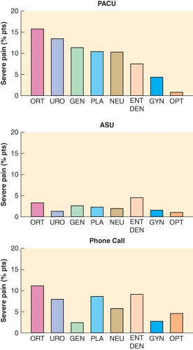 Figure 54.1. Percentage of patients experiencing severe pain in the postanesthesia care unit (PACU), the ambulatory surgery unit (ASU), and during phone call follow-up at 24 hours. ORT, orthopedics; URO, urology; GEN, general; PLA, plastics; NEU, neurology; ENT, ear, nose, throat; DEN, dental; GYN, gynecology; OPT, ophthalmology. (Reprinted from: Chung F, Ritchie E, Su J. Postoperative pain in ambulatory surgery. Anesth Analg. 1997;85:808, with permission.) |
pain, especially during emergence. Administration of parenteral analgesics or sedatives can acutely worsen hypoventilation, airway obstruction, or hypotension, causing sudden deterioration. Evaluating orientation, the level of arousal, and cardiovascular or pulmonary status usually identifies such patients.
Surgical pain can be effectively treated with intravenous opioids as part of a planned analgesic continuum that begins prior to the induction of surgical anesthesia and continues throughout the postoperative course. Sufficient analgesia is the end point, even if large doses of opioids are necessary in tolerant patients. Short-acting opioids are useful to expedite discharge and minimize nausea in ambulatory settings,19 although duration of analgesia can be a problem. During intravenous titration of opioids, assess for incremental respiratory or cardiovascular depression. Disadvantages of intramuscular administration include larger dose requirements, delayed onset, and unpredictable uptake in hypothermic patients. Oral and transdermal analgesics have a limited role in the PACU but are helpful for ambulatory patients after PACU discharge. Rectal analgesics are sometimes useful in small children.
Perioperative use of cyclooxygenase-2 inhibitors has decreased because of adverse cardiovascular events. These events have led to the withdrawal of most of this class of drug with the exception of celecoxib, which has shown to reduce opioid requirements and the incidence of opioid adverse events.20 The concerns surrounding the negative cardiac side effects have made the overall appropriateness of this therapy more complicated. Nonselective nonsteroidal anti-inflammatory drugs such as ibuprofen or acetaminophen are also effective, especially when administered orally before surgery. Intravenous acetaminophen, now available in the United States can also lower opioid requirements. Preoperative administration likely augments the overall level of analgesia rather than offering a substantial preemptive advantage.21 Ketorolac is an effective analgesic and anti-inflammatory that lowers opioid requirements, although the possibility of hemorrhage due to its antiplatelet properties can limit its use. Although intraoperative use has not been associated with increased bleeding, preoperative oral administration has some evidence of postoperative hemorrhage. Ketorolac might also decrease ischemic events in patients with coronary artery disease through analgesic and antiplatelet actions. Use of clonidine to supplement analgesia is effective but can cause hypotension. Agonist–antagonist analgesics offer little advantage. Interventions such as repositioning, reassurance, or extubation also help minimize discomfort.
Other analgesic modalities provide pain relief in and beyond the PACU.22 Intravenous opioid loading in the PACU is important for smooth transition to intravenous patient-controlled analgesia. Injection of opioids into the epidural or subarachnoid space during anesthesia or in the PACU yields prolonged postoperative analgesia in selected patients.23,24 Nausea and pruritus are troubling side effects, and immediate or delayed ventilatory depression can occur related to vascular uptake and cephalad spread in cerebrospinal fluid. Nausea should resolve with antiemetics, whereas pruritus and ventilatory depression often respond to naloxone infusion. Addition of local anesthetic or clonidine enhances analgesia and decreases the risk of side effects from epidural opioids, although local anesthetics add risk of hypotension or motor blockade. Epidural analgesia is effective after thoracic and upper abdominal procedures and helps wean obese patients or those with chronic obstructive pulmonary disease (COPD) from mechanical ventilation. Whether epidural analgesia improves surgical outcomes remains debatable.
Continuous flow catheters with pressure delivery systems of local anesthetics have been used intrawound to reduce pain and opioid requirements, increase patient satisfaction, and reduce length of hospital stay.25 These same delivery systems have been safely used with continuous peripheral nerve catheters for in-hospital as well as outpatient use.26,27 With the use of ultrasound-guided techniques for catheter placement, appropriately selected outpatients can safely receive the pain control benefits of regional anesthesia.9 However, extensive written and oral postoperative instructions must be provided, with systems in place for 24-hour access by patients for catheter-related complications.
Placement of long-acting regional analgesic blocks reduces pain, controls sympathetic nervous system activity, and often improves ventilation.22 After shoulder procedures, interscalene block yields almost complete pain relief with only moderate inconvenience from motor impairment. Paralysis of the ipsilateral diaphragm can impair postoperative ventilation in patients with marginal reserve, although the impact is small in most patients.28 Suprascapular nerve block might be an alternative to avoid this potentially serious side effect. Percutaneous intercostal or paravertebral blocks reduce analgesic requirements after thoracic, breast, or high abdominal incision, although beneficial effects on postoperative pulmonary function are questionable. Transversus abdominis plane (TAP) blocks are effective for lower abdominal surgeries as well as those innervated by the ilioinguinal and iliohypogastric nerves. Caudal analgesia or paravertebral blocks can also be effective in children after inguinal or genital procedures, whereas infiltration of local anesthetic into joints, soft tissues, or incisions decreases the intensity of pain. Other modalities, such as guided imagery, hypnosis, transcutaneous nerve stimulation, music, massage, or acupuncture, have limited utility for surgical pain but may provide a positive patient experience.
Use of patient-controlled analgesia, spinal opioids, or neural blockade mandates anticipation of risk beyond the PACU. The plan for extended postoperative analgesia should be prepared before induction of surgical anesthesia, and then orient the anesthetic and PACU care toward that plan. These plans should be in agreement with the patient, surgeon, and anesthesiologist. If one analgesic modality proves inadequate, take particular care when implementing a second technique.
Fear, anxiety, and confusion often accentuate postoperative pain during recovery, especially after general anesthesia. Titration of an intravenous sedative such as midazolam attenuates this psychogenic component, although analgesic requirements may increase slightly because benzodiazepines interact with γ-aminobutyric acid receptors. It is important to distinguish between requirements for analgesia and for anxiolysis. Opioids are poor sedatives and anxiolytics, whereas benzodiazepines are poor analgesics. However, when opioid dose appears larger than what might be anticipated as what the patient should require, one should consider the possibility that anxiety is playing a large role in the dysphoric event in the PACU.
Discharge Criteria
When possible before discharge from postoperative care, each patient should be sufficiently oriented to assess his or her physical condition and be able to summon assistance. Airway reflexes and motor function must be adequate to maintain patency and prevent aspiration. One should ensure that ventilation and oxygenation are acceptable, with sufficient reserve to cover minor deterioration in unmonitored settings. Blood pressure, heart rate, and indices of peripheral perfusion should be relatively constant for at least 15 minutes and appropriately near baseline. Achieving normal body temperature is not an absolute requirement, but there should be resolution of shivering. Acceptable analgesia
must be achieved and vomiting appropriately controlled. Patients should be observed for at least 15 minutes after the last intravenous opioid or sedative is administered to assess peak effects and side effects. If regional anesthetics have been administered, longer observation could be appropriate. One should monitor oxygen saturation for 15 minutes after discontinuation of supplemental oxygen to detect hypoxemia and then assess likely complications of surgery (e.g., bleeding, vascular compromise, pneumothorax) or of underlying conditions (e.g., hypertension, myocardial ischemia, hyperglycemia, bronchospasm). One should also document a brief neurologic assessment of orientation, eye signs, facial symmetry, and extremity movement and review results of diagnostic tests. If these generic criteria cannot be met, postponement of discharge or transfer to a specialized unit is advisable. There is no demonstrable benefit from a mandatory minimum duration of PACU care.
must be achieved and vomiting appropriately controlled. Patients should be observed for at least 15 minutes after the last intravenous opioid or sedative is administered to assess peak effects and side effects. If regional anesthetics have been administered, longer observation could be appropriate. One should monitor oxygen saturation for 15 minutes after discontinuation of supplemental oxygen to detect hypoxemia and then assess likely complications of surgery (e.g., bleeding, vascular compromise, pneumothorax) or of underlying conditions (e.g., hypertension, myocardial ischemia, hyperglycemia, bronchospasm). One should also document a brief neurologic assessment of orientation, eye signs, facial symmetry, and extremity movement and review results of diagnostic tests. If these generic criteria cannot be met, postponement of discharge or transfer to a specialized unit is advisable. There is no demonstrable benefit from a mandatory minimum duration of PACU care.
Table 54-2. Two Most Commonly Used Postanesthesia Care Unit Discharge Criteria Systems | ||||||||||||||||||||||||||||||||||||||||||||||
|---|---|---|---|---|---|---|---|---|---|---|---|---|---|---|---|---|---|---|---|---|---|---|---|---|---|---|---|---|---|---|---|---|---|---|---|---|---|---|---|---|---|---|---|---|---|---|
| ||||||||||||||||||||||||||||||||||||||||||||||
Postoperative Evaluation
The Centers for Medicare and Medicaid Services (CMS) have instituted compliance policies for those entities that participate in the Medicare and Medicaid programs. The policy for postanesthesia follow-up requires a written documentation that is performed by an individual that is qualified to administer anesthesia no later than 48 hours postprocedure. The time frame starts as soon as the patient arrives to the recovery area or ICU. The evaluation should be performed only after the patient has sufficiently recovered from anesthesia to be able to participate such as answer questions or perform simple tasks. The postanesthesia evaluation must contain the following elements:
Respiratory function, including respiratory rate, airway patency, and oxygen saturation
Cardiovascular function, including pulse rate and blood pressure
Mental status
Temperature
Pain
Nausea and vomiting
Postoperative hydration.
Cardiovascular Complications
Congestive heart failure is epidemic in our ever-aging population. The outpatient cardiology services have an expanding armamentarium of new inotropic/vasodilator therapy, devices, and interventions that allow patients to compensate for their congestive heart failure. One should know not only the ejection fraction but the activities of daily living, exercise tolerance, and other risk indices. The ejection fraction is only an estimate of the fractional shortening of the myocardial actin and myosin fibrils. Although it is a useful estimate of severity of impairment, one is struck by how stable some patients may be with a large dilated heart contracting at a 15% ejection fraction. They are compensated but have little reserve. The potential problems of bleeding, volume shifts, and respiratory compromise in the PACU could quickly cause decompensation. There are also no absolute numbers with regard to fluid restriction. The usage of transesophageal echocardiography revolutionized cardiac anesthesia. It, along with transthoracic echo, may be of great use in the PACU. Within a very few minutes a puzzling hypotensive situation might be explained by an echocardiogram. In the fast-paced dynamic environment of the PACU, placing a pulmonary artery (PA) catheter may give useful information, but may also take valuable time away from patient triage and treatment. The echocardiogram allows rapid viewing of myocardial contractility, regional wall motion, volume status, and valvular dysfunction.
The PACU has in the recent history taken on a new role in some hospitals. Cardiac surgical care is pushing toward “early extubation” or “fast-tracking.” In years past, especially when a “cardiac anesthetic” involved very large dosages of semisynthetic opioids that obligated patients to ≥24 hours of ventilation, the ICU was the standard place for all postoperative heart patients. Today, there is no such entity as a cardiac anesthetic. Balanced anesthetic techniques are used most often. Those who write about early extubation have pushed the limits from 24 hours all the way to extubation of patients on the operating table. Series are available with few if any reintubation catastrophes or events when this technique is practiced with good teams. The natural extension is to establish some highly specialized PACUs that function as step-down or short-term ICUs. In a study of 85 prospective patients31 undergoing “off-pump” coronary artery bypass graft procedures, the patients were extubated in 12 ± 2 minutes after the chest was closed. They were then taken to a special part of the PACU where they were monitored for a number of hours (up to 480 minutes in some situations). Patients were then either discharged to the cardiac floors or sent to an ICU. Of the 85 patients in this study, only 4 failed the PACU stay and had to be admitted to an ICU. Bradycardia was the cause for failure in three cases and one there was one case of myocardial infarction. Two patients later returned to the ICU from the cardiac ward; there was one case of atrial fibrillation and another case of myocardial infarction. During the same time 304 patients who were not undergoing off-pump coronary artery bypass graft surgery were admitted to the cardiac ICU. The cost for PACU stay was $5,140.00 less than for an ICU-admitted patient. Although this study seems quite favorable, the two groups of patients were not comparable.
Studies from the mid-to-late 1990s looking at high-risk vascular and thoracic surgery patients showed that they could each be adequately cared for in an adequately staffed and prepared PACU.32 The conclusion was that a hospital could well improve its patient throughput by putting more resources into expanded PACU care and not so much into ICU services. Several nursing reviews are available to give input as to how to structure such new units.33,34
Anesthesiology services are in increased demand throughout most hospitals. The PACU will likely need to prepare to care for those patients or to staff “ectopic” sites. In the evoked potential laboratories, for example, ablation procedures for dysrhythmias and the newer “mini-Maze” procedures may require care in the PACU. Automated implantable defibrillators are placed in hybrid suites, operating rooms, or catheterization laboratories. Now there is the possibility of percutaneous valve replacements as well as some hybrid and percutaneous coronary revascularization procedures. If these patients require deep sedation or general anesthesia, the patient will also require PACU care.
The cardiac patient is the common patient today. The new procedures and pressure to ever streamline operating room care is pressuring the PACU to become more and more a cardiac mini-ICU. The smart PACU medical director and hospital administrator will see that with targeted resources, patients may well be safely cared for in a more cost-effective manner with quicker throughput by using a PACU approach.
Postoperative Pulmonary Dysfunction
Mechanical, hemodynamic, and pharmacologic factors related to surgery and anesthesia impair ventilation, oxygenation, and airway maintenance.35 Heavy smoking, obesity, sleep apnea, severe asthma, and COPD increase the risk of postoperative ventilatory events.36 Preoperative pulmonary function testing has limited predictive value for postoperative complications,37 perhaps with the exception of postoperative bronchospasm in smokers.38
Inadequate Postoperative Ventilation
In PACU patients, mild respiratory acidemia due to atelectasis and decreased minute ventilation is expected; thus, elevated PaCO2 does not necessarily indicate inadequate postoperative ventilation. Inadequate ventilation should be suspected when (1) respiratory acidemia occurs coincident with tachypnea, anxiety, dyspnea, labored ventilation, or increased sympathetic nervous system activity; (2) hypercarbia reduces the arterial pH below 7.30; or (3) PaCO2 progressively increases with a progressive decrease in arterial pH.
Inadequate Respiratory Drive
During early recovery from anesthesia, residual effects of intravenous and inhalation anesthetics blunt the ventilatory responses to both hypercarbia and hypoxemia. Sedatives augment depression from opioids or anesthetics and reduce the conscious desire to ventilate (a significant component of ventilatory drive).
Hypoventilation and hypercarbia can evolve insidiously during transfer and admission to the PACU. Although effects of intraoperative medications are usually waning, the peak depressant effect of an intravenous opioid given just before transfer occurs in the PACU. Coincident depression of medullary centers that regulate the sympathetic nervous system can blunt signs of acidemia or hypoxemia such as hypertension, tachycardia, and agitation, concealing hypoventilation. Patients might communicate lucidly and even complain of pain while experiencing significant opioid-induced hypoventilation. A balance must be struck between an acceptable level of postoperative ventilatory depression and a tolerable level of pain or agitation. Patients with abnormal CO2/pH responses from morbid obesity, chronic airway obstruction, or sleep apnea are more sensitive to respiratory depressants.39 Risk for apnea after anesthesia in preterm infants depends on type of anesthetic, postconceptual age, and preoperative hematocrit. Preterm infants should be monitored for at least 12 hours (see Chapter 41). Children with active or recent upper respiratory infection are more prone to breath-holding, severe cough, and arterial desaturations below 90% during recovery, especially if they have a history of reactive airway disease or secondhand smoke exposure or have undergone intubation and/or airway surgery.40 If hypoventilation from opioids is excessive, forced arousal and careful titration (20 to 40 μg at a time) of intravenous naloxone reverses respiratory depression without affecting analgesia. Flumazenil (0.1 mg titrated to effect up to 1.5 mg) directly reverses depressant effects of benzodiazepines on ventilatory drive but is usually unnecessary.
The abrupt diminution of a noxious stimulus (e.g., tracheal extubation, placement of a postoperative block) may promote hypoventilation or airway obstruction by altering the balance between arousal from discomfort and depression from medication. Intracranial hemorrhage or edema sometimes presents with hypoventilation, especially after posterior fossa craniotomy. Bilateral carotid body injury after endarterectomy can ablate peripheral hypoxic drive. Chronic respiratory acidemia from COPD alters CNS sensitivity to pH and makes hypoxic drive dominant, but hypoventilation from supplemental oxygen rarely occurs.
Increased Airway Resistance
High resistance to gas flow through airways increases work of breathing and CO2 production. If inspiratory muscles cannot generate sufficient pressure gradients to overcome resistance, alveolar ventilation fails to match CO2 production and progressive respiratory acidemia occurs.
In postoperative patients, increased upper airway resistance is caused by obstruction in the pharynx (posterior tongue displacement, change in anteroposterior and lateral dimensions from soft-tissue collapse), in the larynx (laryngospasm, laryngeal edema), or in the large airways (extrinsic compression from hematoma, tumor, or tracheal stenosis). Weakness from residual neuromuscular relaxation,41 myasthenia gravis or myasthenic syndromes can contribute, but it is seldom the primary cause of airway compromise. If the airway is clear of vomitus or foreign bodies, simple maneuvers such as improving the level of consciousness, lateral positioning, chin lift, mandible elevation, or placement of an oropharyngeal or nasopharyngeal airway usually relieve obstruction. A nasopharyngeal airway is better tolerated when the patient has functional gag reflexes. Acute extrinsic upper airway compression (e.g., an expanding neck hematoma) must be relieved.
During emergence, stimulation of the pharynx or vocal cords by secretions, blood, foreign matter, or extubation can generate laryngospasm.42 Laryngeal constrictor muscles occlude the tracheal inlet and reduce gas flow. Patients who smoke or are chronically exposed to smoke have irritable airway conditions, have copious secretions, or have undergone upper airway surgery are at higher risk.35,40 Laryngospasm can usually be overcome by providing gentle positive pressure (10 to 20 mm Hg continuous) in the oropharynx by mask with 100% O2. Prolonged laryngospasm is relieved with a small dose of succinylcholine (e.g., 0.1 mg/kg) or deepening sedation with propofol. An intubating dosage of succinylcholine should not be used to break postoperative laryngospasm, especially if the alveolar partial pressure of oxygen (PAO2) is decreased by hypoventilation. As little as 5 to 10 mg of succinylcholine can break the laryngospasm. Unless assisted ventilation is provided, declining PAO2 causes serious hypoxemia before spontaneous ventilation resumes43 (Fig. 54-2). If the functional residual capacity (FRC) is abnormally reduced, the decreased volume of O2 available in the lungs accelerates the development of hypoxemia. Severe laryngeal obstruction can occur secondarily because of hypocalcemia after parathyroid excision.
Soft-tissue edema worsens airway obstruction, especially in children and adults recovering from procedures on the neck. Nebulized vasoconstrictors like epinepherine help somewhat, but steroids have little effect acutely. Patients with C1 esterase inhibitor deficiency can develop severe angioneurotic edema after even slight trauma to the airway. Pathologic airway obstruction (e.g., severe edema, epiglottitis, retropharyngeal abscess, encroaching tumors) might require emergency tracheal intubation, but airway manipulation is dangerous because minor trauma from intubation attempts can convert a marginal airway into a total obstruction. Judgment by the individual anesthesiologist regarding timing, patient status, available equipment along with airway management skills all play a part of the decision as to where, when, and how to intubate. Sedatives or muscle relaxants used
to facilitate intubation can worsen obstruction by compromising the patient’s volitional efforts to maintain the airway and by eliminating spontaneous ventilation. Equipment and personnel necessary for emergency cricothyroidotomy or tracheostomy should be available. Needle cricothyroidotomy using a 14-gauge intravenous catheter or a commercially available kit permits oxygenation and marginal ventilation until the airway is secured, especially if jet ventilation with 100% oxygen is used.
to facilitate intubation can worsen obstruction by compromising the patient’s volitional efforts to maintain the airway and by eliminating spontaneous ventilation. Equipment and personnel necessary for emergency cricothyroidotomy or tracheostomy should be available. Needle cricothyroidotomy using a 14-gauge intravenous catheter or a commercially available kit permits oxygenation and marginal ventilation until the airway is secured, especially if jet ventilation with 100% oxygen is used.
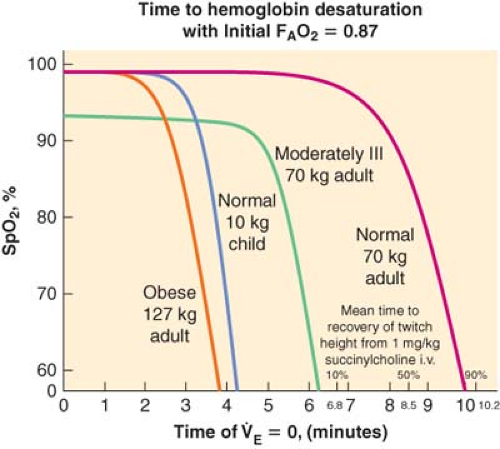 Figure 54.2. Rate of SpO2 decline after onset of apnea. (Reprinted from: Benumof JL, Dagg R, Benumof R. Critical hemoglobin desaturation will occur before return to an unparalyzed state following 1 mg/kg intravenous succinylcholine. Anesthesiology. 1997;87:979, with permission.) |
Related posts:


Full access? Get Clinical Tree


