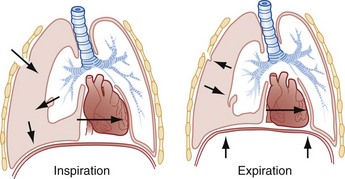
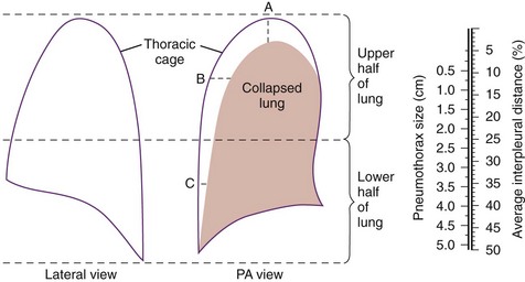
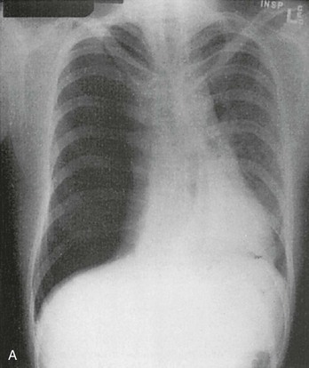
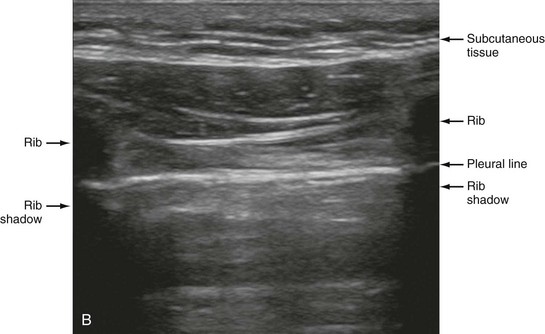
Pleural Disease

Chapter 77


Pleural Disease


Pleural disease is commonly encountered in the emergency department (ED). Presentations range in severity from asymptomatic pleural effusion to tension pneumothorax. This chapter reviews the two most common nontraumatic pleural problems: spontaneous pneumothorax and pleural inflammation with effusion. Pleural space problems associated with trauma are discussed in Chapter 45, and the approach to a patient with pleuritic chest pain in Chapter 26.


Spontaneous Pneumothorax


Perspective


Under normal conditions, the visceral and parietal pleurae lie in close apposition, with only a potential space between them. Pneumothorax is defined as the presence of free air in the intrapleural space. A spontaneous pneumothorax occurs in the absence of any external precipitating factor, either traumatic or iatrogenic. Primary spontaneous pneumothorax occurs in individuals without clinically apparent lung disease. Secondary spontaneous pneumothorax arises in the context of an underlying pulmonary disease process.


The incidence of primary spontaneous pneumothorax is approximately 15 cases per 100,000 population per year among men and 5 cases per 100,000 population per year among women.1,2 Primary spontaneous pneumothorax typically occurs in healthy young men of taller than average height. Factors associated with primary spontaneous pneumothorax include cigarette smoking and changes in ambient atmospheric pressure. Familial patterns suggest an inherited propensity in some cases of primary spontaneous pneumothorax. Mitral valve prolapse and Marfan syndrome are associated with spontaneous pneumothorax in the absence of clinically apparent lung disease.


Approximately one third of spontaneous pneumothoraces occur in the context of underlying pulmonary disease (Box 77-1). The incidence of secondary spontaneous pneumothorax is three times higher in men. The most common condition associated with secondary spontaneous pneumothorax is chronic obstructive pulmonary disease (COPD), which accounts for nearly 70% of cases. Patients with severe COPD (e.g., with forced expiratory volume in 1 second below 1 L) are at highest risk. The incidence of spontaneous pneumothorax among patients hospitalized for emphysema is 0.8% and for asthma 0.3%.


Spontaneous pneumothorax occurs in approximately 2% of patients with acquired immunodeficiency syndrome, almost always in the setting of Pneumocystis jiroveci (previously known as Pneumocystis carinii) pneumonia.


Malignancy is another common cause of secondary spontaneous pneumothorax. The occurrence of spontaneous pneumothorax in a patient with known malignancy should suggest lung metastases. In developing countries, tuberculosis and lung abscess remain leading causes of secondary spontaneous pneumothorax.


Catamenial pneumothorax is a rare condition in which recurrent spontaneous pneumothorax occurs in association with menses (typically within 72 hours of onset). Although it is termed thoracic endometriosis syndrome and often responds to ovulation-suppressing medications, the exact cause of catamenial pneumothorax is uncertain.


Spontaneous pneumothorax is rare in childhood. The principles of diagnosis, imaging, treatment, and surgical management for pediatric primary spontaneous pneumothorax are similar to those for adult pneumothorax.1


Pathophysiologic Principles


Normally, intrapleural pressure is negative (less than atmospheric), fluctuating from −10 mm Hg to −12 mm Hg during inspiration to approximately −4 mm Hg during expiration. Intrabronchial and intra-alveolar pressures are negative during inspiration (−1 to −3 mm Hg) and positive during expiration (+1 to +3 mm Hg). The alveolar walls and visceral pleura form a barrier that separates the intrapleural and intra-alveolar spaces and maintains the pressure gradient. If a defect occurs in this barrier, air enters the pleural space until either the pressures equalize or the communication seals.


With the loss of negative intrapleural pressure in one hemithorax, the ipsilateral lung collapses. A large pneumothorax results in restrictive ventilation impairment, with reduced vital capacity, functional residual capacity, and total lung capacity. Shunting of blood through nonventilated lung tissue may result in acute hypoxemia, although over time this effect is mitigated by compensatory vasoconstriction in the collapsed lung.


In tension pneumothorax, the alveolar-pleural defect acts as a one-way valve, allowing air to pass into the pleural space during inspiration and trapping it there during expiration (Fig. 77-1). This trapping leads to progressive accumulation of intrapleural air and increasingly positive intrapleural pressure, causing compression of the contralateral lung with asphyxia and worsening hypoxia. Intrapleural pressure exceeding 15 to 20 mm Hg impairs venous return to the heart. If the situation is allowed to progress, cardiovascular collapse and death ensue.


In primary spontaneous pneumothorax, disruption of the alveolar-pleural barrier occurs when a subpleural bulla (or bleb), typically located at the lung apex, ruptures into the pleural space. Subpleural bullae are found in almost all patients who undergo surgical treatment for primary spontaneous pneumothorax and are identified on computed tomography (CT) of the chest in 90% of cases. The cause of these bullae may be related to degradation of elastic fibers within the lung and an imbalance in the protease-antiprotease and oxidant-antioxidant systems.2


In the case of secondary spontaneous pneumothorax, the underlying lung disease weakens the alveolar-pleural barrier. In patients with P. jiroveci pneumonia, the cytotoxic effects of repeated episodes of inflammation lead to bullous and cystic changes. In patients with COPD, chronic exposure to cigarette smoke results in the development of large, thin-walled bullae that are at an increased risk for rupture. Other factors, including increased intrabronchial and intra-alveolar pressures generated by bronchospasm and coughing, also play a role.


Clinical Features


Symptoms of primary spontaneous pneumothorax typically begin suddenly while the individual is at rest. Ipsilateral chest pain and dyspnea are the most common symptoms. At the outset, the pain is typically “pleuritic” in nature (i.e., often described as sharp and made worse with deep inspiration), but it often evolves over time into a dull, steady ache. Although patients frequently describe shortness of breath, extreme dyspnea is uncommon in the absence of underlying lung disease or tension pneumothorax. Cough is present in a few individuals. Occasionally, patients are asymptomatic or have only nonspecific complaints. Patients may wait several days before they seek medical attention, and a significant number delay presentation for 1 week or more. Without treatment, symptoms often resolve spontaneously within 24 to 72 hours, although the pneumothorax is still present.


Physical findings tend to correlate with the degree of symptoms. A mild sinus tachycardia is the most common physical finding. With a large pneumothorax, decreased or absent breath sounds with hyper-resonance to percussion may be present. Other classic signs include unilateral enlargement of the hemithorax, decreased excursion with respirations, absent tactile fremitus, and inferior displacement of the liver or spleen. Absence of any or all of these findings does not exclude pneumothorax, however, and a chest radiograph should be obtained when pneumothorax is suspected.


With tension pneumothorax, signs of asphyxia and decreased cardiac output develop. Tachycardia (often >120 beats/min) and hypoxia are common. Hypotension is a late and ominous finding. Distention of the jugular veins is common but may be difficult to detect. Displacement of the trachea to the contralateral side is classically described but is an uncommon finding, usually occurring only in the immediately preterminal phase of the pneumothorax, if at all. Its absence should not be considered evidence that a tension phenomenon is not present.


In patients with significant underlying lung disease, pneumothorax manifests differently. Because of poor pulmonary reserve, dyspnea is nearly universal, even when the pneumothorax is small, and symptoms tend not to resolve on their own. Physical findings, such as hyperexpansion and distant breath sounds, often overlap considerably with the underlying lung disease, making the clinical diagnosis difficult. For this reason the diagnosis of pneumothorax should be considered whenever a patient with COPD experiences an exacerbation of dyspnea.


Although suggested by the patient’s history and physical examination, the diagnosis of pneumothorax is generally made with the chest radiograph. The classic radiographic appearance is that of a thin, visceral pleural line lying parallel to the chest wall, separated by a radiolucent band devoid of lung markings. The average width of this band can be used to estimate the size of the pneumothorax with a fair degree of accuracy (Fig. 77-2). In general, however, it is more reasonable simply to characterize the pneumothorax as small, moderate, large, or total. The estimated size of the pneumothorax and the patient’s clinical status can be useful in guiding management decisions.


Tension pneumothorax is a clinical diagnosis, and delay in treatment to obtain radiographic confirmation is inadvisable. When the diagnosis of tension pneumothorax is not apparent clinically and a chest radiograph is obtained, the classic appearance is one of complete lung collapse with gross distention of the thoracic cavity on the affected side and shift of mediastinal structures across the midline (Fig. 77-3A). In patients with underlying pulmonary disease, however, pleural adhesions and lack of lung elasticity may mask the fact that a pneumothorax is under significant positive pressure.


When pneumothorax is suspected but not seen on a standard chest radiograph, an expiratory film may be obtained. Theoretically, the volumes of the lungs and the chest cavity are reduced during expiration so that the relative size of the pneumothorax is enhanced. Although expiratory films are occasionally helpful in identifying a small apical pneumothorax, their routine use does not improve diagnostic yield. In critically ill patients for whom only a supine chest radiograph can be obtained, the finding of a “deep sulcus” (i.e., a deep lateral costophrenic angle) can suggest the presence of pneumothorax on that side (see Fig. 77-3A).


Special care should be taken in viewing the chest radiographs of patients with underlying lung disease. In patients with COPD, the relative paucity of lung markings makes pneumothorax more difficult to detect. At the same time, giant bullae may simulate the radiographic appearance of pneumothorax. A clue to differentiating a pneumothorax from a giant bulla is that the former tends to run parallel to the chest wall, whereas the latter tends to have a more concave appearance. When the diagnosis is unclear, CT can differentiate between the two entities.


Chest CT is considered the gold standard for the diagnosis of pneumothorax; however, it requires that patients be stable enough for transport. Although portable chest radiography is convenient and commonly used, it has poor overall sensitivity.3 Point-of-care thoracic ultrasound is a rapid and accurate diagnostic aid performed and interpreted by the clinician at the bedside.4,5 Bedside thoracic ultrasound is more sensitive than a supine chest radiograph for the diagnosis of pneumothorax and avoids ionizing radiation.6,7 In addition to routine use for the diagnosis of both traumatic and spontaneous pneumothorax, thoracic ultrasound can be used to evaluate for postprocedural pneumothorax and for the diagnosis of occult pneumothorax in critically ill patients.8,9 In normal lung the closely opposed visceral and parietal pleura create the appearance of shimmering or sliding of the pleural interface during respiration. Assessment for pneumothorax is optimally performed with a high-frequency linear probe to most accurately visualize the lung sliding at the pleural line (see Fig. 77-3B). In supine patients, air rises; scanning a few interspaces on the anterior chest wall along the midclavicular line can effectively evaluate the most likely areas for pneumothorax. Extending the examination to the lateral chest wall can help identify the “lung point” or boundary between normal lung and pneumothorax and is highly specific for the detection of pneumothorax.10 Visualization of lung sliding at the pleural line effectively rules out pneumothorax in the area being scanned. A small pneumothorax may not be detected with ultrasound, and false-positive results can occur in patients with blebs, pleural scarring, or severe acute respiratory distress syndrome (ARDS) or in single-lung intubation.


The differential diagnosis of pneumothorax includes numerous conditions associated with chest pain and dyspnea. Among the most important is pulmonary embolism, which may manifest in similar fashion with unilateral pleuritic chest pain. Most pleural-based processes (pneumonia, embolism, tumor) have characteristic radiographic findings. Rarely, pneumothorax may mimic an acute myocardial infarction with electrocardiographic changes simulating an acute injury pattern.


Spontaneous pneumomediastinum is a closely related clinical entity, diagnosed by the presence of subcutaneous emphysema and the finding of mediastinal air on chest radiography. In contrast to spontaneous pneumothorax, spontaneous pneumomediastinum typically occurs during exertion, particularly after a strenuous Valsalva maneuver. Most cases of spontaneous pneumomediastinum occur in the absence of known underlying disease and have a benign course. Secondary causes of pneumomediastinum (e.g., Boerhaave’s syndrome) are more serious, and treatment is aimed at the underlying disorder.


Spontaneous hemopneumothorax is a rare but potentially serious condition that occurs when collapse of the lung is associated with rupture of a vessel in a parietopleural adhesion. The clinical presentation is similar to that of spontaneous pneumothorax but may be accompanied by symptoms and signs of hemorrhagic shock. Treatment entails large-caliber tube thoracostomy to evacuate the pleural space, reexpand the lung, and tamponade bleeding.


Pneumothorax has a readily available, highly reliable confirmatory diagnostic test (i.e., chest radiography). Absence of a pneumothorax on chest radiography should prompt a search for an alternative diagnosis.


Management


Whether in the field or in the ED, if the clinical circumstances suggest tension pneumothorax, treatment should be initiated without awaiting further cardiovascular compromise or definitive diagnosis by chest radiography. As soon as tension pneumothorax is suspected, the pleural space should be decompressed. This decompression may be accomplished by insertion of an intravenous catheter or by immediate tube thoracostomy, depending on the availability of equipment and the expertise of the providers. The diagnosis is confirmed by the hiss of air escaping under positive pressure as the needle or chest tube enters the pleural space. Needle decompression is only a temporizing procedure, and definitive management requires prompt tube thoracostomy. In morbidly obese patients, the needle and catheter may be of insufficient length to reach the pleural space, and a longer needle may be required.


The management of spontaneous pneumothorax has two goals: (1) to evacuate air from the pleural space, and (2) to prevent recurrence. Pursuit of the latter goal extends beyond the realm of the ED but may influence the initial approach to management. Therapeutic options for treatment of pneumothorax range from simple observation or aspiration with a catheter to video-assisted thoracoscopic surgery or thoracotomy. Decisions should be individualized and consider several factors, including size of the pneumothorax, severity of signs, presence of underlying pulmonary disease, other comorbidities, history of previous pneumothoraces, patient reliability, degree and persistence of the air leak, and available follow-up monitoring.


For otherwise healthy, young patients with a small primary spontaneous pneumothorax (i.e., <20% of the hemithorax), and minimal symptoms, observation alone may be appropriate. The intrinsic reabsorption rate ranges from 1 to 2% per day, a rate that is accelerated by a factor of 4 with the administration of 100% oxygen. By lowering the alveolar partial pressure of nitrogen, supplemental oxygen increases the rate at which air diffuses across the pleural-alveolar barrier. Admission to the hospital generally is not required for a patient with a small pneumothorax and no hemodynamic disturbance or hypoxia. Although there are no evidence-based guidelines, we recommend observing these patients in the ED for a 4- to 6-hour period. This observation period also can occur in an ED-based observation unit. A repeat chest radiograph obtained before discharge confirms that there is no interval worsening in the size of the pneumothorax. Discharged patients should be able to return to an ED if they worsen and should undergo telephone or in-person follow-up with a primary care provider in 24 to 48 hours. Air travel and underwater diving must be avoided until the pneumothorax has completely resolved. Patients who are not able to follow up with a primary care physician in the desired interval are advised to return to the ED for follow-up evaluation.


For primary spontaneous pneumothoraces that are larger in size (i.e., ≥20% of the hemithorax), aspiration with an intravenous catheter may be attempted. If 6 hours after aspiration the chest radiograph shows no reaccumulation of the pneumothorax, the catheter is removed and the patient can be discharged home, with the same caveats that apply to patients managed with observation alone.


Although there is no universal agreement on the optimal treatment of patients with a first episode of primary spontaneous pneumothorax, data suggest that aspiration may be equally effective as chest tube drainage and clearly causes much less patient discomfort.11 Advantages of simple aspiration include low morbidity, lack of invasiveness, and overall cost savings, with reported rates of successful outcome ranging from 45 to 71%.12 Success is less likely when the patient is older than 50 years or the volume of air aspirated exceeds 2.5 L, suggesting a continuing air leak. If aspiration fails to reexpand the lung fully, the catheter can be attached to a water-seal device or to a one-way Heimlich valve and managed like a small-caliber chest tube.


Most recurrent spontaneous pneumothoraces should be managed with tube thoracostomy because less invasive approaches (i.e., observation or simple aspiration) are associated with significantly lower success rates.13 Similarly, patients who have respiratory distress, have tension physiology, or are likely to require mechanical ventilation should undergo tube thoracostomy to reexpand the lung definitively. Also, if there is significant pleural fluid (hemothorax or hydrothorax), tube thoracostomy is required. Finally, tube thoracostomy may be considered in uncomplicated cases of primary spontaneous pneumothorax either as a first-line intervention or after a less invasive approach (i.e., observation or simple aspiration) fails.


For most primary spontaneous pneumothoraces, placement of a small-caliber (7-14F) tube is generally sufficient because air leakage tends to be minimal. Small-caliber tubes are easy to insert, are well tolerated by patients, and leave only a small scar after removal. Complications associated with small-caliber tubes include kinking, malposition, inadvertent removal, occlusion by pleural fluid or clotted blood, and large persistent air leaks. For secondary spontaneous pneumothorax, a standard size (20-28F) thoracostomy tube is recommended. When there is detectable pleural fluid or an anticipated need for mechanical ventilation, a larger tube size (≥28F) is required.


After insertion, the tube is attached to a water-seal device and left in place until the lung has reexpanded fully and the air leak has ceased. A Heimlich valve, which consists of a one-way flutter valve covered in transparent plastic, can be used in place of a water-seal device and allows unhindered ambulation. Specific complications associated with the use of a Heimlich valve include accidental disconnection and occlusion by fluid.


Routine application of suction neither increases the rate at which the lung reexpands nor improves patient outcome and is no longer recommended. The use of suction (with a pressure of 20 cm H2O) is reserved for situations in which the lung fails to reexpand after drainage through a water-seal device or Heimlich valve for 24 to 48 hours.


In most cases, chest tube management requires hospital admission, although outpatient management of spontaneous pneumothorax with a small-caliber tube and Heimlich device also is reasonable in a stable, responsible patient with good follow-up availability. Common complications of chest tube placement include incorrect placement, pleural infection, and prolonged pain. Reexpansion pulmonary edema and reexpansion hypotension are rare occurrences after rapid evacuation of large pneumothoraces.

< div class='tao-gold-member'>

Jul 26, 2016 | Posted by in ANESTHESIA | Comments Off on Pleural Disease

Full access? Get Clinical Tree

Get Clinical Tree app for offline access