 Emergent venous access for fluid resuscitation or drug infusion if alternative peripheral or central access is either unattainable or contraindicated
CONTRAINDICATIONS
 Absolute Contraindications
 Major blunt, long-bone fracture or penetrating trauma proximal to site of cutdown
 Relative Contraindications
 Suspected proximal vascular injury (in the extremity or inferior vena cava)
 Overlying infection, burn, or skin damage at the site of cutdown
 Coagulopathy
RISKS/CONSENT ISSUES
 Pain (local anesthesia can be given)
 Local bleeding and hematoma
 Infection (sterile technique will be utilized)
 General Basic Steps
 Analgesia
 Isolate
 Stabilize
 Cannulate
 Secure
LANDMARKS
Greater saphenous vein is most easily accessible 1 to 2 cm anterior and 1 to 2 cm superior to the medial malleolus. The vein may be palpable in a nonhypotensive patient (FIGURE 26.1).
SUPPLIES
 Povidone–iodine or chlorhexidine solution
 Anesthetic supplies: 1% lidocaine, 1 vial, 10-mL syringe, 22- or 25-gauge needle
 No. 10 and no. 11 scalpel blades
 Curved Kelley hemostat
 Catheter-over-the-needle, 16 or 18 gauge
 Needle driver
 Silk sutures (3-0 and 4-0), nylon suture (4-0)
 Iris scissors
 Intravenous tubing and injectable sterile saline
 Wound dressing supplies
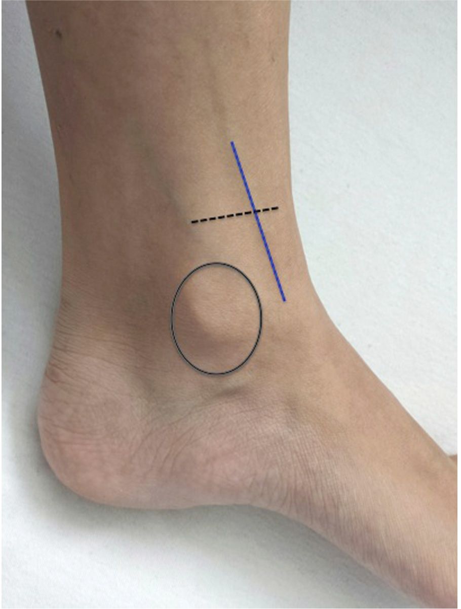
FIGURE 26.1 Saphenous vein runs vertically 2 cm anterior to the medial malleolus of the ankle. The ideal site of cutdown is 2 cm superior to the medial malleolus (dotted lines).
TECHNIQUE
 Patient Preparation
 Extend and externally rotate lower extremity
 Immobilize if needed, especially in children
 Sterilize entire ankle with povidone–iodine solution and drape
Note: Unless immediate emergent access is warranted, universal precautions (cap, eye shields, mask, sterile gown and gloves) should be worn.
 Analgesia
 Use 25- or 27-gauge needle to anesthetize skin and subcutaneous tissue with 1% lidocaine
 Isolate: FIGURES 26.2A and B
 Make a skin incision transversely over saphenous vein landmark
 Apply traction to skin on either side of the incision to expose subcutaneous tissue
 Dissect the subcutaneous tissue using a curved hemostat with the tip facing downward, parallel to the course of the vein
 After exposing the vein, pass the hemostat under the vein, turn the tip upward, and spread to isolate the vein above the hemostat
 Stabilize: FIGURE 26.2C
 Pass two 3-0 or 4-0 silk suture ties under the vein using the curved hemostat
 Clamp each tie with hemostats, one proximally and the other distally
 Distal suture may be tied to ligate the vessel. This decreases bleeding, but also sacrifices the vessel.
 Apply traction on each tie, thereby lifting the vessel and exposing the anterior surface of the vein
 Cannulate: FIGURES 26.2D and E
 With the tip of no. 11 scalpel blade make a flap incision to the anterior surface of the vein, approximately one-third the diameter of the vein
 A vein pick may be used to elevate the flap
 Carefully advance the catheter through the incision
 Flush the catheter with saline solution and attach to intravenous line
 Secure Catheter: FIGURE 26.2F
 Tie the proximal suture around the vein and the IV catheter to secure in place
 Cut the ends of the proximal and distal ties
 Suture the catheter to the skin and close the incision using 4-0 nylon sutures
 Cover skin insertion site with antibiotic ointment and sterile dressing
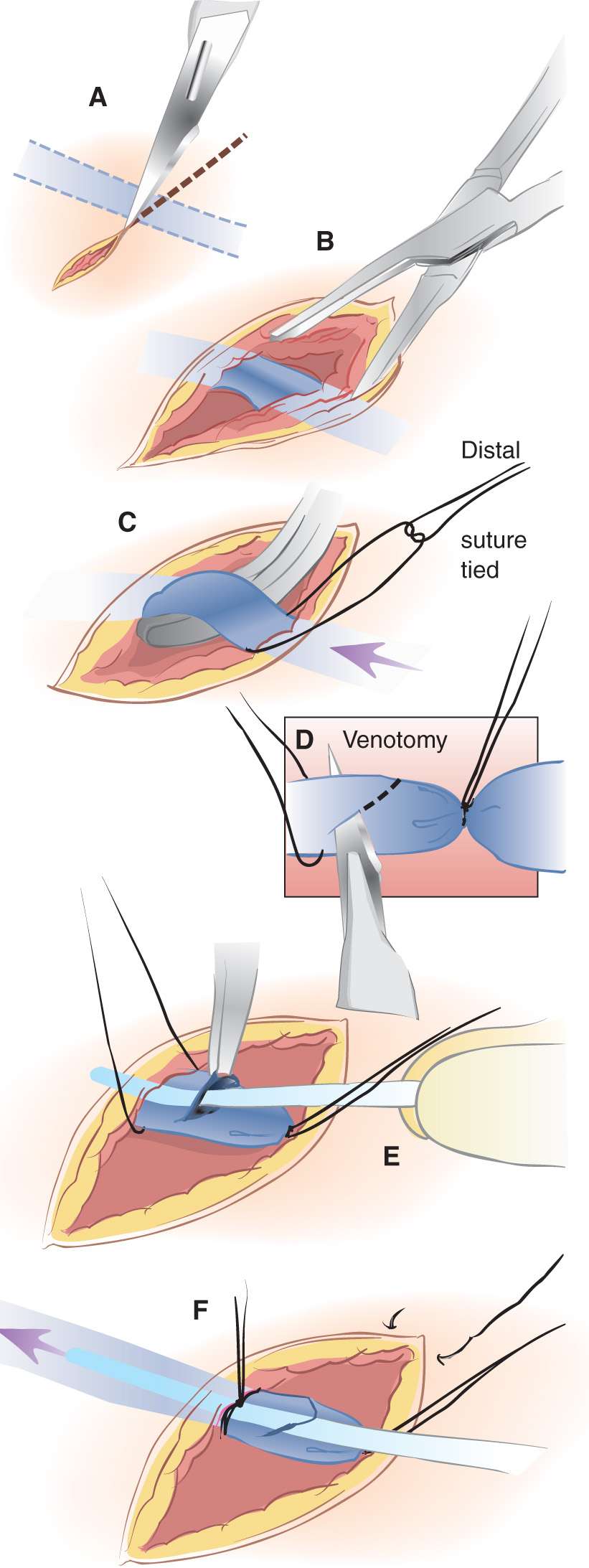
FIGURE 26.2 Procedures for venous cutdown catheterization. A: A transverse incision is made 2 cm anterior and superior to medial malleolus. The incision should extend into the subcutaneous tissue but not deep enough to potentially lacerate the vein. B: The vein is isolated using blunt dissection. A suture is passed around the vein and cut to give two ligatures. C: The distal suture is tied and used to stabilize the vessel. D: Venotomy is performed to allow insertion of the catheter. Alternatively the surgeon may choose to insert the catheter over a needle without performing a venotomy, in a manner similar to percutaneous catheterization. E: The catheter is inserted into the vein. Placement within the vessel is confirmed by aspirating blood or infusing fluid. F: The proximal suture is tied and the wound is closed and dressed. (From Vinci RJ. Venous cutdown catheterization. In: Henretig FM, King C, eds. Textbook of Pediatric Emergency Procedures. Philadelphia, PA: Williams & Wilkins; 1997:284, with permission.)



Full access? Get Clinical Tree


