Mechanical Ventilation
Rarely, some patients who are recovering from anesthesia may need some form of mechanical ventilation in the PACU. Various techniques such as positive end-expiratory pressure (PEEP), continuous positive airway pressure (CPAP), and synchronized intermittent mandatory ventilation (SIMV) are used to improve the respiratory status of the patient. Table 28.2 gives the terminology of the common ventilatory modes.
Positive End-Expiratory Pressure
PEEP is a technique that can be used with invasive ventilation to help prevent collapse of the alveoli during the expiratory phase of ventilation, to increase the lung’s functional residual capacity, and to reduce the amount of physiologic shunting. PEEP also increases the PaO2, which usually enables the FiO2 to be reduced, thus lessening the chances of oxygen toxicity. In patients with preexisting obstructive lung disease, PEEP should be used cautiously because it can overexpand relatively normal alveoli. When PEEP is used in such circumstances, the dead space increases and occasionally causes a decrease in the PaO2 and an increase in the PaCO2.
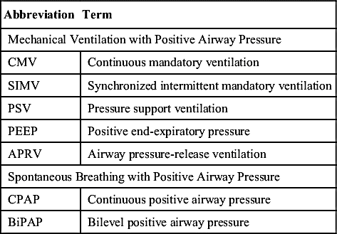
When a patient receives PEEP therapy, hemodynamic status should be monitored because this ventilatory technique decreases venous return and may cause a decrease in cardiac output, especially in the patient with hypovolemia. In some instances, the reduced cardiac output can cause a decrease in systolic blood pressure. Other parameters to be monitored are vital signs, skin perfusion, and urine output.
Continuous Positive Airway Pressure
CPAP applies a constant pressure during both inspiration and expiration and helps to keep the lungs expanded. The patient breathes out against increased pressure as high as 10 to 20 cm H2O. The lung performs at a larger, more inflated volume, thereby increasing the functional residual capacity and decreasing the tendency to atelectasis. CPAP is a technique that can be used for weaning a patient from a ventilator or applied to the patient who is spontaneously breathing but needs pressure support. Patients with histories of obstructive sleep apnea may benefit from use of CPAP in the postanesthesia care unit.7 When CPAP is used, the patient should be monitored for tachypnea, tachycardia, increases in blood pressure, dysrhythmias, or generalized distress, which should be reported to the physician, if detected.
Bilevel Positive Airway Pressure
Bilevel positive airway pressure (BiPAP) is another form of noninvasive positive-pressure ventilation that provides continuous high-flow positive airway pressure that cycles between two pressure levels, a higher positive pressure (IPAP, inspiratory positive airway pressure) and a lower positive pressure (EPAP, expiratory positive airway pressure). Gas exchange improves with BiPAP because of an increase in alveolar ventilation. Monitoring is similar to that for CPAP.
Synchronized Intermittent Mandatory Ventilation
Synchronized intermittent mandatory ventilation (SIMV) provides both mandatory breaths at a set rate and allows the patient to take spontaneous breaths in between. It allows the patient to control the inspiratory time and the size of the spontaneous tidal volumes depending on patient effort and muscle strength, lung compliance, airway resistance, and whether pressure support is present to increase spontaneous tidal volume delivery. If the patients are apneic, they will receive the mandatory breaths at a set rate. If they take a breath in between mandatory breaths, they will receive a pressure support assisted breath. If they take a breath near when the mandatory breath is ready to deliver a breath, the ventilator will “synchronize” delivery of the mandatory breath with the patient effort.
Nursing Responsibilities
All PACU nurses must be familiar with the specific types and modes of operation of ventilators used in the area (Fig. 28.1; see Table 28.2); however, some nursing responsibilities remain the same regardless of mechanical ventilator. The following list discusses these responsibilities.
1. Ascertain that the patient’s lungs are ventilated with frequent observation of the chest for bilateral synchronous and equal expansion and by listening for bilaterally present and equal breath sounds.
2. Check the airway frequently for complete patency. See that the patient’s ventilator system is free of significant leaks by listening for air gurgling in the upper airway during ventilation and comparing the exhaled volume with the tidal volume set on the ventilator.
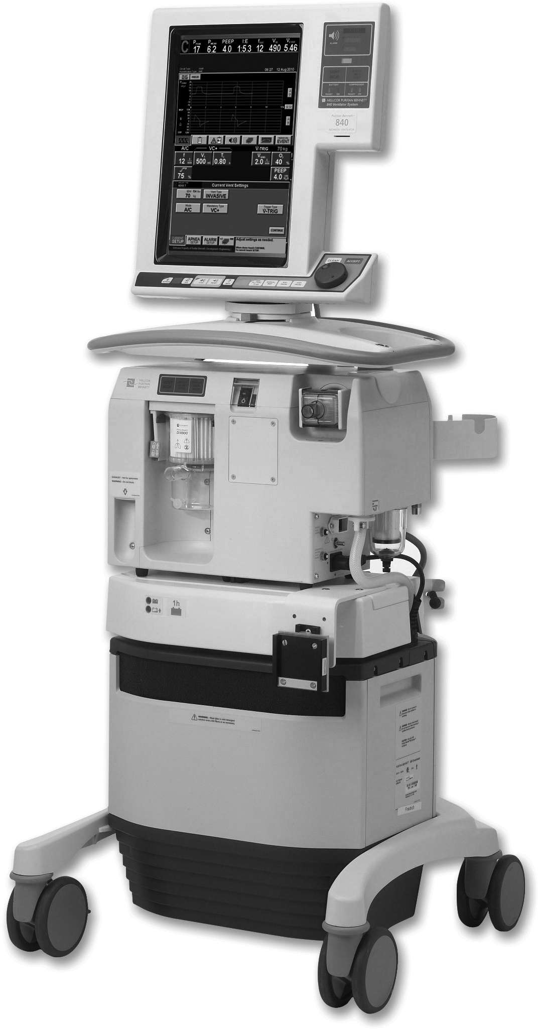
FIG. 28.1 840 Ventilator system. Volume and pressure ventilator used either to assist or to control a patient’s respirations and offer SIMV, BiLEVEL, and spontaneous modes. (©2016 Medtronic. All rights reserved. Used with the permission of Medtronic.)
3. Ensure that the cuff is never overinflated. Measure the cuff pressure and maintain it between 20 to 30 cm H2O.
4. Empty the ventilatory hoses frequently of excess water from condensation.
5. Ensure that proper humidification is delivered to the patient by noting the presence of water droplets in the ventilator hoses.
6. Check the humidifier and fill frequently to ensure proper humidification.
8. Position the patient’s endotracheal or tracheostomy tube so there is never any pull.
9. Perform the tracheostomy wound care as needed during the postanesthesia phase.
10. Ascertain frequently that all alarms on the ventilator are on, set appropriately, and functioning properly.
The observations and checks of mechanical devices often seem simple and routine but are an important part of nursing the patient who requires mechanical ventilation. Ideally, all these checks, along with measured parameters of the patient’s respiratory status, should be recorded either on a flow sheet attached to the patient’s bed or to the ventilator or electronically as defined by the facility. In most facilities, respiratory therapists maintain and manage the ventilator in the PACU. The respiratory therapist is available to assist the nurse caring for the patient who requires a ventilator and to complete the appropriate documentation of ventilator and patient status.
Suctioning
When large amounts of secretions accumulate that cannot be handled effectively with coughing, suctioning must be instituted to assist the patient in clearing air passages.
Oral and Nasal Suctioning
Suctioning the nose and mouth is simple and safe. This procedure is commonly used to assist patients in eliminating secretions when they have not yet regained full consciousness and cannot spit out secretions. The catheter should be soft and pliable. The technique should be clean but need not be strictly sterile. A Yankauer or tonsil suction tip can be used to remove oral secretions from the mouth and over the tongue; however, care must be used to avoid breaking or chipping the teeth.
Tracheal Suctioning
Tracheal suctioning can be performed through the nose via endotracheal tube or through a tracheostomy tube (Fig. 28.2). Tracheal suctioning must be accomplished atraumatically with aseptic technique. A selection of sterile suctioning catheters in a variety of sizes should be kept at the bedside of every patient in the PACU along with sterile gloves and sterile water or normal saline solution. The catheter chosen for suctioning should not have an external diameter that exceeds by one third the internal diameter of the tube to be suctioned. Most commonly, a 14 or 16 F size is used for adult patients. The catheter must not completely occlude the trachea or endotracheal tube.


Full access? Get Clinical Tree


