 Paraphimosis (foreskin trapped proximal to coronal sulcus) is a urologic emergency, and reduction is always indicated when the condition is present
CONTRAINDICATIONS
 None
None
RISKS/CONSENT ISSUES
 Pain (local anesthesia will be given)
 Local bleeding
 Infection (sterile technique will be used)
 Scarring at site of incision/dilation (though definitive treatment will likely include circumcision, removing the scarred tissue)
 Damage to glans penis and urethral meatus
 General Basic Steps
If manual reduction fails to restore normal anatomy, proceed to assisted manual reduction; if that fails as well, proceed to phimotic ring incision. A urologist should be contacted if manual reduction fails, and will need to be actively involved if emergency circumcision is required because of reduction failure.
 Patient preparation
 Anesthesia
 Manual reduction
 Assisted manual reduction (FIGURE 37.1)
 Phimotic ring incision
TECHNIQUE
 Patient Preparation
 Position: Supine with legs slightly abducted
 Consider light procedural sedation as well
 Anesthesia
 A topical anesthetic may be all that is necessary
 Apply viscous lidocaine or eutectic mixture of local anesthetics (EMLA) to the inner layer of the foreskin
 Also serves as a lubricant
 If further anesthesia is necessary, progress from local, to dorsal nerve block, and, finally, to penile ring block
 See chapter 36 for details and pictures of penile anesthesia
 When providing local anesthesia with lidocaine (without epinephrine), be sure to infiltrate into the constricting ring as well
 Manual Reduction
 Using your hands or an elastic bandage, manually compress the glans and foreskin for 3 to 5 minutes to remove as much edema as possible
 Place both thumbs on the glans penis and apply slow, steady pressure while using your other fingers just proximal to the phimotic ring to pull the foreskin over the glans penis (FIGURE 37.2)
 Successful reduction occurs when the phimotic foreskin is reduced back to normal position over the glans
 If unsuccessful, proceed to assisted manual reduction methods
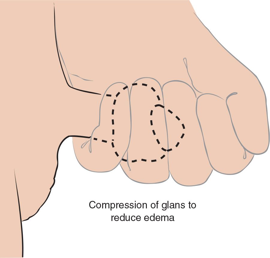
FIGURE 37.1 Technique for compression of glans to reduce edema and allow for manual reduction of paraphimosis. (From Green M, Strange GR. Paraphimosis reduction. In: Henretig FM, King C, eds. Textbook of Pediatric Emergency Procedures. Philadelphia, PA: Williams & Wilkins; 1997:1008, with permission.)



Full access? Get Clinical Tree


