Physical neuromatrix, including peripheral and central nervous systems plus fascial network:
• Emotional
• Cognitive
• Memory
• Mind
Physical Laser Beams
We live with and through a dynamically fluctuating nervous system, one which has a marvelously complex functioning in terms of pain transmission [13, 18, 19]. To briefly review, eudynia starts with stimulation of chemical, mechanical, or temperature nociceptors in the periphery [33, 34]. Via transduction, an action potential is created, and this electrical signal is conducted to the spinal dorsal horn (Fig. 1.1). Here, a complex series of interactions occur, with Aδ and C fibers working to enhance the signal strength, while Aβ fibers and descending inhibitory fibers work to inhibit the signal, and all of this interaction receives an additional excitatory influence from the glial cells [35, 36] within the dorsal horn. Once the dorsal horn interactions reach a final summation of factors, the remaining signal is then transmitted via the spinothalamic and spinoparabrachial tracts to the brain (Fig. 1.2). The spinothalamic transmission is delivered to the thalamus and is processed on to the contralateral somatosensory cortex. The spinoparabrachial transmission is processed through the hippocampus, amygdala, and onward to the prefrontal cortex, ACC, and other areas of the brain bilaterally (Fig. 1.3) [13, 26, 27, 37–40].
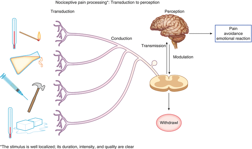
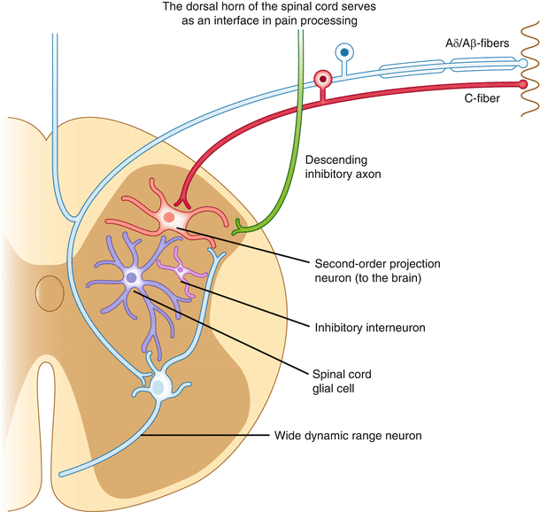
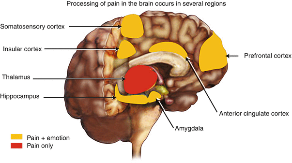
Fig. 1.1
Nociceptive pain processing. Transduction to perception
Fig. 1.2
The dorsal horn of the spinal cord serves as an interface in pain processing
Fig. 1.3
Processing of pain in the brain occurs in several regions
Intensification of the laser beam being generated by the periphery or spinal cord can occur via sensitization of the peripheral nociceptors, which increases the intensity of the signal reaching the spinal cord [12]. At the spinal level, we can experience recruitment of new nociceptive inputs or even non-nociceptive fibers (as with Aβ fibers) when the signals are strong enough. When enough stimulation has occurred via root input or dorsal horn sensitization, the spinal cord can go into “automatic” mode, where it no longer needs peripheral input to fire. Thus, we can wind up with very strong and enduring laser beams from the physical generators below the brain [13, 38, 39, 41].
Within the brain, adding further to this complex process which happened in the dorsal horn, an area of the caudate contains TANS (tonically active neurons). It is in this area of the brain where a confluence of signals from the hippocampus (memory), our emotions (amygdala), and our cognitions are processed, with the resultant signals being sent to the globus pallidus and up to the motor cortex. Thus, messages that come from areas of our brain that are overlapping and interacting with pain signals can incorporate cognitive, emotional, and memory inputs that have a direct effect on our motor system as well as our sensory system [6]. This provides an understanding to the concept that our brain processing is geared to result in “action” and is responsive to our sensory perceptions such as pain. We do not experience sensory perceptions for the sake of experiencing alone [6 14 22, 42, 43].
In addition to the neural network, physical input to our pain hologram can be strongly influenced by the fascial system, described as the organ system of stability and mechano-regulation [44], and wherein lies ten times more sensory nerve endings than in muscle [45, 46]. Fascia lines all body parts, as John Barnes puts it: “fascia is a tough connective tissue that spreads throughout the body in a three-dimensional web from head to foot functionally, without interruption” [2]. The purpose of fascia is to maintain body shape and keep organs in their proper positions, as well as resist mechanical stresses from any source such as trauma and inflammation [47]. This function of fascia is best understood through the construct of tensegrity, a concept that explains the relationship between skeleton, tensional forces of the fascia, contractility of muscle, and hydrostatic pressure of fascial compartments [48]. Restrictions of the fascia have been found to cause limitations in movement and pain which can have non-dermatomal referral patterns [49, 50], thus demonstrating that the physical input to our pain hologram from fascia is not by activation of peripheral nociceptors. While often ignored in evaluating a person’s pain perception, fascial contribution to pain has been demonstrated in such diverse problems as Achilles tendinopathy [51], plantar fasciitis [52], systemic lupus erythematosus [53], and acute compartment syndrome of the upper extremity [54].
Researchers have demonstrated an energy transmission system throughout fascial planes. This energy wave is faster than neural transmission and is very nicely visually demonstrated by Guimberteau [55]. Body memory of past events or trauma (physical, emotional, or sexual) [3] can be stored in the fascia, similarly to neural storage, and this stored memory can interrupt the smooth flow of energy via the fascial system [2, 55–57]. We do know that traumatic events are processed by the person immediately after they happen. However, what is not totally processed at that time is stored in cellular body memory traces that become like a three-dimensional photo (although the storage is in energy units and not pictures) and incorporates all contingencies of that event, placing them in “storage” at an unconscious level. Barnes has found that a part of these stored memory traces in the fascial network are positionally dependent [2]. Through Barnes’ unwinding technique, bodily positions that can replicate the same body part position at the time of the trauma can release and make conscious the stored unconscious memories and allow the person to finish working through that event [2].
Other therapeutic applications that take advantage of this fascial network input to pain are being used for surgical anesthesia and post-op pain control. Fascial iliaca compartment block has been used in fractured neck of femur [58], hip arthroplasty [59], and this same block has been shown to reduce emergence agitation in children having thigh surgery [60]. One study cited has replaced epidural anesthesia with fascial anesthesia in prostatectomies [61]. Although the fascial network is not processed via the peripheral nociceptors, there is some recent animal research to indicate some dorsal horn activity via the fascial network in addition to fascial activation itself, and this article concludes that fascial input is a significant contributing force to painful syndromes [62].
Emotional Laser Beams
We are all familiar with the fact that people with persistent pain frequently have complaints of anger, anxiety, depression, and sometimes fear attached to their pain experience. These reactions have been categorized into phasic, acute, and chronic by Craig, with phasic and acute representing anticipatory fear and relief, while chronic represents depression, fear, anger, disgust, social distress, guilt, subservience, resignation, and abandonment [63]. The question then arises as to whether these emotional complaints are representative of primary psychological illnesses or are they part and parcel of normal and/or dysfunctional brain activity relative to pain perception? Do people living with maldynia have multiple illnesses or was Osler correct to have us think of “one person, one disease”?
Perhaps we can make more sense of this question by looking at brain function. Brain areas significantly involved in emotion in the brain include the amygdala, hippocampus, lateral hypothalamus, caudate, anterior cingulate cortex (ACC), supraorbital cortex, and prefrontal cortex [13, 37]. These same areas have been well documented in depression, anxiety, obsessive-compulsive disorder, and fear among others [4, 6, 11]. Interestingly, if we look at the affective dimension of pain, according to Price and the Rome article, these same brain areas are the ones involved [14, 15, 28]. Thus, what we are beginning to identify is that pain perception and emotional problems share some of the same “brain railroad tracks.” The brain doesn’t “know” what it is doing, it just does. Therefore, if the brain is utilizing the same tracks for two different types of perception, it cannot tell which train is riding that track at any given time, nor does it care. In fact, two trains can use the same tracks at the same time, and by so doing, they can signal a “go” to each other. This mixed signaling helps us understand why people in pain, especially those with maldynia, will report that stressful or depressing events can exacerbate their pain. In our holographic analogy, this would be equivalent to adding strength to some of the laser beams making up our pain hologram, by non-painful inputs. This could be likened to “recruitment” in the spinal cord, where we intensify a pain signal through recruitment of non-nociceptive fibers. (Spinal recruitment is a lower level route to add strength to the “physical” pain laser beam being fed into our hologram.)
The medical literature supports the reverse concepts to also be a frequent occurrence; that is, psychiatric patients with affective disorders often have pain as a symptom of their affective disorder. Phillips and Hunter identified an increased prevalence and intensity in tension-type headaches in a psychiatric population compared to the general population [64]. Melzack and Katz have discussed that stressful events have been associated with angina pectoris, ulcers, rheumatoid arthritis, painful menses, ulcerative colitis, and regional enteritis [20].
In addition, some psychiatrists have taken the position that pain is no more than a symptom of psychiatric disease and is not a disease unto itself. We believe the distinction is better conceived by understanding perception rather than disease states. For example, Romano and Turner have written that approximately 50 % of all patients with pain and depression develop the two “disorders” simultaneously [65]. In view of brain imaging studies and our current understanding of overlapping brain areas in pain and depression, it makes sense that some patients may experience pain and depression simultaneously, while others may feel one or the other first. If both perceptions are utilizing the same brain areas and reinforcing each other, then it becomes easier to understand why depression could stimulate a pain perception, pain could stimulate depression, or both could start together. Remember, the brain is the only part of the body that can “perceive,” and since the brain only “does,” without understanding, then any combination of perceptions can take place if the same areas of the brain are being utilized for them.
Cognitive Laser Beams
Marcus Aurelius [66] once said:
If you are distressed by anything external, the pain is not due to the thing itself, but to your estimate of it. THIS you have the power to revoke at any time.
Our brain is structured such that the most primitive areas, in terms of development, are lower in location. Our cortex has been described as having evolved to be sitting on top of the older brain. Thus, the areas that are so highly integrated into the affective dimension of pain, as well as much of the pain areas associated with maldynia, are for the most part sub/lower cortical. As mentioned above, the TANS is the location where the cognitive areas meet the emotional areas, which have had input already from memory. Like so many of our sensory perceptions, the lower brain takes charge rather than the logical inputs we are capable of. It is often said that in most any issue between emotions and logic, the emotions will win out, that is, we will default to the “heart.” This is another way of saying that decisions and responses, unless consciously influenced, will include “unconscious” influences that are more emotionally driven. Perlmutter and Villodo discuss the role of prefrontal cortex in reasoning and creative thinking and how changes in prefrontal functioning can lead to a “dysregulation” of the balance necessary for optimum brain function [67].
This “default” system can often lead us into difficulty. For example, when a person takes a medication for pain relief, the feeling of relief (“feels good”) can easily lead us into the behavior of “if one feels good, then two or three must be even better.” And hence, we can wind up with a patient developing significant adverse medication reactions by their instinctual (unconscious) desire to be pain free. Too much NSAID, acetaminophen, antidepressant, antiepileptic, etc., can produce physical harm to the body. Too much controlled substance can produce adverse bodily reactions and/or behaviors that result in legal trouble as well. The ultimate expression of this “action without thinking” response can be the development of pseudoaddiction, where the perception of pain relief is the desired goal, and our behaviors can mimic those of someone with a true addictive disorder. One person’s actions are driven by the desire to relieve pain, while the person with an addictive disorder demonstrates behavior driven by the need to get high and, further into the disease, by the need to avoid crashing and experiencing withdrawal. The behaviors may be very similar on the part of those two different people, with lack of demonstrable control in following prescription directions, drug seeking behaviors, actually placing themselves in harm’s way at times, lying to those around them in order to obtain more medications, etc. (see Chap. 6 on Addictive Disorders and Pain). What we experience in situations where the lower brain centers are controlling our responses are cognitive rationalizations, that is, “if one pill works, two is better” as a way to justify our desire to have less pain. This kind of cognitive laser beam is one in which the cognition follows the affective dimension rather than lead it.
Various issues regarding the cognitive input to pain perception have been described [4]. These contributions have looked at such issues as the roles of language descriptors [68], emotion and attitudes [69], culture and attention [70], ethnicity [71], gender differences [72], and age differences (see Chap. 19, Pain in the Elderly Population) [73]. The literature also supports the significance of the affective dimension through the cognitive inputs [23]. These contributions from mind and cognition support Schwartz’s description of processing the affective, memory, and cognitive processes through the TANs, described earlier.
Conversely, when cognitive-behavioral therapies are utilized in treating pain or other problems such as depression or OCD, the success of the patient depends on their ability, through much practice, to have the cognitive abilities of the higher cortex take charge and present alternative “thinking” to the patient. This change allows for a reframing of thoughts and a refocusing of attention as well as the consequent behaviors away from the “painful” thoughts, that is, “I hurt,” “I am suffering,” “I will never be able to enjoy family picnics again,” etc [4, 74]. These examples are of our cognitive system in a passive mode (default system). The alternatives, through cognitive-behavioral approaches, would be to assertively place into consciousness such concepts as “this pain has no beneficial meaning to me, therefore, I will focus on the love I have for antique cars and review some pictures of old convertibles now,” or “since this pain is meaningless to me, I choose to breathe deep ten times and allow my body to feel the flow of positive energy course through me,” etc. Utilizing our higher cortical powers assertively, then, allows us to change the “default” system by building in new neuroplastic patterns. We literally can control our lives by creating the new set of railroad tracks we want our train to utilize and set up the switching mechanisms by practicing, until the new track becomes our “default.” This becomes active mode for our cognitive inputs [6].
Thus, the cognitive contribution to pain lasers can be positive or negative and can be minimal when in passive mode (old default), or when in active mode, the cognitive input has the potential, in many instances, to become the most powerful influence to overcome adverse emotional reactions [23]. As we will see below, the cognitive force from our mind through our cortical thinking brain can become a valuable source of positive neuroplastic retraining of our brain. Cognitive-behavioral processes have been shown to be the most effective in helping people with maldynia to restore their functional status and maximize their abilities to take charge of their lives again [4, 74]. Our developmentally highest level of brain function is often needed to help us deal with our most significant life problems, when our lower brain levels that normally run on automatic default fail us. The paradox is that it so often requires professional help to teach us how to utilize these higher level approaches to alter our life experiences, which are our perceptions (see Chap. 7).
Another example of how we can utilize our higher cognitive power to defeat pain is through hypnosis. Rainville et al. have demonstrated that if we use hypnosis to alter the affective-motivational dimension of pain first, there is often a reduction in the sensory-discriminative pain dimension that follows it [75]. However, this approach makes changes in brain function through the lower centers which mediate the affective dimension of pain and does not involve the somatosensory cortex. On the other hand, the reverse is not true. If we utilize hypnosis to alter the sensory-discriminative dimension of pain first, the process does involve the somatosensory cortex, but even if we alter the pain intensity, the affective dimension (the “yuck”) doesn’t change [75]. Thus, how we build our new railroad tracks and which “switchers” we utilize can have a rather dramatic effect on the “retraining” process of the brain (see Chap. 9).
Memory Laser Beams
Memory in humans is a complex process, which involves multiple inputs to go from immediate memory to long-term memory. Memory is made in the body cells [7, 56] as well as in the brain, but it is not made in pictures. It is made in mnemonics, with different memory storage for each part of the memory. For example, the memory for a traumatic event (painful or not) will have memory traces for the event itself, the place it happened, the smells involved, the sounds heard, the sights seen, the emotions perceived, the thoughts associated with the event, our judgment of what has happened, etc. The memory is made and stored according to events and patterns. The memory may or may not remain in our conscious awareness, but long-term memory is permanent [76, 77].
Brain areas involved in memory involve left prefrontal, temporal, and parahippocampal cortices. The level of activation of these areas predicts which memory becomes long term or not [78, 79]. The hippocampus and amygdala relate to emotional memory (remember the TANS) via NMDA and dopamine, and long-term potentiation in the hippocampus may underlie learning and memory [76, 77, 80]. Prefrontal cortex is involved with object identity, spatial locations, memory and coding, and analysis of the meaning of items [81–83]. Prefrontal cortical involvement increases as the semantic complexity rises [78, 79]. Thus, we can begin to see how memory becomes entwined with the confluence of brain patterning involved in pain perception. Through the TANS, the feed-in of memory, emotions, and meaning all meld together. When the input is of sufficient intensity, memory will be made [83].
When we want to actively recall a memory, such as “what did I eat for lunch today,” our brain must activate these brain areas, pull up the mnemonic for each part of that hologram for “lunch,” and converge them all to produce the three-dimensional holographic picture in my mind’s eye of lunch today. This will include all my senses, including taste, color, food presentation, sounds in the room where lunch was eaten, the conversation that took place, who was there, the temperature of the food and room, how much comfort or discomfort was involved both physically and emotionally, etc.
Thus, memory is made in parallel for the events and for the emotional and other components [15, 28]. Hence, when we have to converge multiple mnemonics to produce our pain perception hologram, the mnemonic for any portion of the pain (sensory-discriminative, affective, memory, or both) can be overloaded by previous memory mnemonics for that particular quality of the pain [4, 84]. If the affective component is overloaded, we can see the limbically augmented pain syndrome (LAPS) described by Rome and Rome. Thus, memory, and sensitization of the memory system, can result in augmentation of the pain perception. This accounts for why sufferers of persistent pain often have a more frequent history of trauma compared to the general population.
Returning to our great Roman emperor, Marcus Aurelius [66], we can again quote him:
As for pain, a pain that is unbearable carries us off; but that which lasts a long time is bearable; the mind retires into itself, and the ruling faculty is not injured. As for the parts which are hurt by the pain, let them, if they can, give their opinion of it.
If we view this memory system through the lenses of the brain areas involved, and realize those same brain areas are involved with the physical, emotional, cognitive, and meaningfulness of any perception, including pain, we can see the genius behind Marcus Aurelius’ two observations about pain perception. He demonstrated a far-reaching wisdom about pain perception, without any scientific knowledge of how accurate his statements have turned out to be in terms of modern investigations into brain functioning.
Mind Control and Mind Laser Beams
Building on the foundation that our mind is something different from our brain, even though it operates through the brain, we can add some very powerful laser beams and control over the entire system through mindfulness.
As Schwartz has described, “quantum theory creates a causal opening for the mind, a point of entry by which mind can affect matter, a mechanism by which mind can shape brain. That opening arises because quantum theory allows intention, and attention, to exert real, physical effects on the brain…” [6].
This same author has brought together the work of many neuroscientists, such as William James, Henry Stapp, and Benjamin Libet in order to demonstrate how the mind can physically affect the brain. It has been demonstrated that a wave of “readiness” energy appears in the brain about 350–550 ms before a motor movement occurs. In addition, the sense of will occurs 150–200 ms prior to a movement. This free will offers an opportunity to make the movement a “go” or “not go” [6]. Stowell has previously described a similar time delay in pain perception [85], and the impact of psychosocial feedback has been investigated in the timing of events by Lee and colleagues [42].
Hence, the understanding of free will becomes a process by which the brain “bubbles up” unconscious thoughts that could lead to action; but free will, as a conscious system, provides an opportunity to screen these bubbling ideas and exert control over which ones are a go or not. It has been proposed that the initiatives that bubble up in the brain are based on the person’s past memories, experiences, values inculcated from society, and present circumstances. Interestingly, studies of brain function in relationship to free will demonstrate that the prefrontal cortex is activated as a primary area. Disorders such as schizophrenia, which is marked by autistic behavior and inactivity, and clinical depression, one symptom of which is lack of initiative, demonstrate a consistently low level of activity in the prefrontal cortex [6].
Related posts:


Full access? Get Clinical Tree


