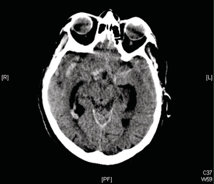
Ian T. Ferguson and Christopher R. Carpenter Department of Emergency Medicine, Washington University School of Medicine, St. Louis, MO, USA Headaches represent about 2.8% of emergency department (ED) visits per year.1 Most headaches are self‐limited, but subarachnoid hemorrhage (SAH) is life threatening and is classically described as a sudden onset “thunderclap headache” reaching maximal severity within seconds. The differential diagnosis of acute onset headaches also includes cerebral vasospasm, hypophyseal apoplexy, venous sinus thrombosis, vascular dissection, cough, exertional and postcoital headache, and migraine.2–6 Migraine‐like headaches are at least 50 times more common than SAH among ED headache patients.7 A meta‐analysis of ED‐based studies provided a pooled point estimate of 7.5% SAH incidence when emergency physicians were concerned about SAH.8 Missed SAH are a substantial medical malpractice risk,9,10 so the evidence‐based diagnostic approach to headache epitomizes emergency medicine via a highly lethal condition without a pathognomonic presentation nested within a high‐volume complaint for which the vast majority do not have a life‐threatening etiology. Approximately 85% of SAH are caused by ruptured aneurysms. Box 46.1 provides the differential diagnosis of nontraumatic SAH. Perimesencephalic bleeds are the second most common etiology. Usually, SAH is diagnosed by noncontrast head computed tomography (CT) (Figure 46.1). The diagnostic accuracy of head CT can be improved by incorporating the suggestions noted in Box 46.2.11 Because early generation CTs missed up to 5% of SAH, classic teaching emphasized the need to proceed to lumbar puncture (LP) to exclude SAH. However, LP infrequently identifies SAH after a negative head CT: the number of LPs required to identify one aneurysmal SAH amenable to surgical intervention after a negative CT performed within 6 hours of symptom onset ranges from 91 to 15,200 LPs.12–14 Test‐treatment threshold analyses provide another way to quantify risks and benefits of a diagnostic test. Two separate test‐treatment analyses for LP when diagnosing SAH provide similar results: only patients with post‐CT SAH probability between 2% and 9% have benefits outweighing risk associated with LP if imaged within 6 hours and lacking any CT evidence of SAH.8,15 Based upon the diagnostic accuracy of CT described below, the pretest probability of SAH would need to exceed 70% for a CT obtained within 6 hours for the posttest probability to fall within the 2–9% range. The 70% pretest probability is far higher than any ED SAH prevalence reported. In addition, LP is not a harmless procedure. Post‐LP headache rates are 6–30%,16 many patients are reluctant to undergo LP,17 and clinicians often do not obtain LPs due to time constraints and low diagnostic yield.18,19 LPs are also often inconclusive. “Traumatic taps” (cerebrospinal fluid [CSF] red blood cells [RBC] secondary to the invasive procedure rather than SAH) occur in about one in six LPs with no universally accepted methods to differentiate SAH CSF RBCs from procedural blood.20 Figure 46.1 Noncontrast head computed tomography (CT) showing an acute subarachnoid hemorrhage. Can history, physical exam, and/or decision aids accurately and reliably distinguish patients at increased or decreased risk of nontraumatic SAH? The American College of Emergency Physician’s (ACEP) 2019 Clinical Policy on “Acute Headache” and a 2016 diagnostic meta‐analysis both emphasize that no element of history or physical exam alone accurately rule in or rule out SAH.8,21 Objective neck stiffness (pooled positive likelihood ratio [LR+] 6.6) and self‐reported stiff neck (pooled LR+ 4.1) most accurately rule in SAH, while the absence of “worst headache of life” (pooled negative likelihood ratio [LR−] 0.24) most accurately rule out SAH. The diagnostic accuracy of history and physical exam are summarized in Tables 46.1 and 46.2, respectively.8 The ACEP Clinical Policy provides a Level B recommendation in favor of the Ottawa SAH Rule to rule out SAH safely without a CT or LP. The Ottawa SAH decision aid (Figure 46.2) identifies patients with suspicion for SAH (which does not mean every ED headache patient) as low‐risk if none of the following features is present: age ≥40, neck pain or stiffness, witnessed loss of consciousness, headache onset with exertion, thunderclap description, or limited neck flexion on examination. The initial validation of the Ottawa SAH decision aid demonstrated a LR− = 0.02 (CI 0.00–0.39).22 Subsequent multisite validation in Canada demonstrated 100% sensitivity and LR− = 0.23 Researchers continue attempts to improve the diagnostic performance of the Ottawa SAH Rule24 or derive new decision aids.25 Theoretically, using the Ottawa SAH would safely decrease CT rates, but prospective implementation studies demonstrate no decrease in CT ordering, although a 14% reduction in LPs and 2.5% lower admission rate has been observed.26 Table 46.1 Predictors of SAH from history Source: Data from [8].
Chapter 46
Nontraumatic Subarachnoid Hemorrhage
Background
Clinical question
Risk factor (number of studies)
Sensitivity, % (95% CI)
Specificity, % (95% CI)
Positive LR (95% CI)
Negative LR (95% CI)
Arrival by ambulance
Pooled accuracy (2)
59 (53–65)
80 (78–81)
2.95 (2.23–3.89)
0.51 (0.44–0.63)
Awoke from sleep
Pooled accuracy (2)
11 (8–16)
82 (80–83)
0.63 (0.45–0.89)
1.09 (1.04–1.14)
Blurred vision
Pooled accuracy (2)
11 (2–28)
95 (92–98)
3.14 (0.31–31.43)
0.85 (0.44–1.63)
Burst or explosion onset
Pooled accuracy (5)
58 (52–64)
47 (45–49)
1.10 (0.75–1.60)
0.88 (0.56–1.38)
Diplopia (1)
0 (0–15)
98 (94–100)
0.96 (0.05–19.33)
1.00 (0.94–1.07)
ED transfer
Pooled accuracy (2)
18 (13–23)
92 (91–93)
2.20 (1.65–2.91)
0.90 (0.85–0.95)
Exertion at onset
Pooled accuracy (5)
29 (24–34)
83 (82–84)
1.46 (1.16–1.83)
1.20 (0.80–1.79)
Family history cerebral aneurysm (1)
0 (0–13)
92 (87–95)
0.22 (0.01–3.54)
1.07 (1.00–1.15)
Female sex
Pooled accuracy (6)
58 (53–62)
41 (39–42)
1.10 (0.93–1.31)
0.93 (0.73–1.12)
Intercourse at onset
Related posts:

Full access? Get Clinical Tree

 Get Clinical Tree app for offline access


