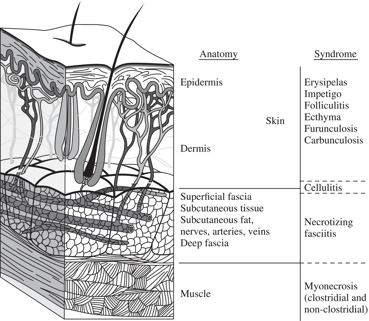
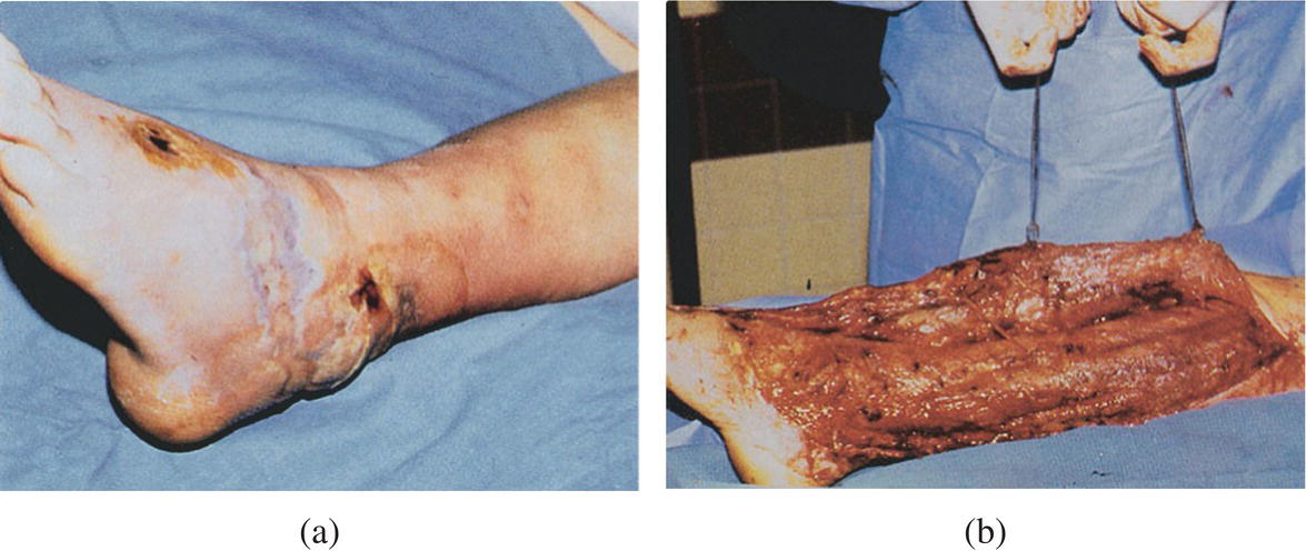
Lucas Oliveira Junqueira e Silva1,2 and Fernanda Bellolio2 1 Department of Emergency Medicine, Hospital de Clínicas de Porto Alegre, Porto Alegre, Brazil 2 Department of Emergency Medicine, Mayo Clinic, Rochester, MN, USA Necrotizing soft tissue infection (NSTI), commonly referred as “necrotizing fasciitis,” is a rapidly progressive infection involving the fascia and subcutaneous tissue. It most commonly affects the extremities. Differentiating necrotizing infection from other skin and soft tissue infections (Figure 25.1) is important in the emergency department (ED). While NSTI is a rare disease, it results in considerable morbidity and mortality. Mortality is estimated at 20% to 30%. NSTI is a surgically treated disease, and early recognition and debridement of necrotic fascia and other involved areas are major determinants of overall outcome (Figure 25.2). A delay in debridement has been associated with poor survival. Early on, NSTI can be difficult to distinguish from other forms of soft tissue infections, such as cellulitis and abscess. Computed tomography (CT), magnetic resonance imaging (MRI), and ultrasound have been used in distinguishing necrotizing fasciitis from other clinical entities. Figure 25.1 Schematic of the different layers of the skin and the corresponding infections associated with each layer. Figure 25.2 (a) A suspected case of necrotizing fasciitis. Left foot shown with oozing wound, dusky skin, and bullae formation. (b) Surgical exploration resulted in extensive debridement. (Hall et al. [1], Reproduced with permission from McGraw‐Hill Education.) Can physical examination be helpful in distinguishing necrotizing soft tissue infection from nonnecrotizing soft tissue infections? Several clinical manifestations are classically described in patients with NSTI, including erythema (without sharp margins), edema that extends beyond the visible erythema, severe pain (“out of proportion”), fever, crepitus, and skin bullae, necrosis or ecchymosis. Some of these findings are often described as “pathognomonic,” which would mean a very high positive predictive value for the diagnosis of NSTI. However, a diagnostic test accuracy systematic review and meta‐analysis found limited evidence for the use of physical examination findings, especially regarding their ability to rule out the diagnosis of NSTI.2 The authors calculated summary estimates for three examination findings: fever, hemorrhagic bullae, and hypotension (Table 25.1). While the pooled positive likelihood ratio was high for hemorrhagic bullae (LR+ 5.97, CI 2.89–12.32) and hypotension (LR+ 9.20, CI 3.87–21.86), these two findings, when absent, had limited ability to change posttest probabilities (pooled negative likelihood ratios of 0.78 and 0.81, respectively). These data illustrate that their absence should not be individually used to rule out the presence of NSTI. The authors also provided a Bayesian‐based clinical reasoning exercise in their discussion, where they reported that a patient with a pretest probability of NSTI of 50% without fever, hemorrhagic bullae, or hypotension, still remains with a posttest probability of 41.3%, 43.9%, and 44.7%, respectively. Table 25.1 Physical examination findings to differentiate necrotizing soft tissue infections from other nonnecrotizing skin and soft‐tissue infections Source: Data from [2]. Physical examination findings may be used to increase the posttest probabilities to levels where no further diagnostic procedures are needed. In other words, these findings may aid clinicians to cross through the threshold for requesting surgical evaluation. For example, a patient with a pretest probability of NSTI of 20% presenting with hypotension (LR+ 9.20) remains with a posttest probability of 70%. This relatively high posttest probability should lead to immediate surgical consultation. On the other hand, if hypotension was absent, the remaining posttest probability would be at 17%, almost not changed from the initial 20%. Can laboratory tests be reliably used in the ED in distinguishing necrotizing soft tissue infection from other nonnecrotizing soft tissue infections? The Laboratory Risk Indicator for Necrotizing Fasciitis (LRINEC) investigators developed a scoring system to differentiate necrotizing fasciitis from other skin and soft tissue infections.3 They derived the LRINEC scoring system in a retrospective cohort of 314 patients and validated it in 140 patients in two teaching hospitals in Singapore. They included 140 patients who had necrotizing fasciitis and 309 patients with severe cellulitis or abscesses. They found that WBC, hemoglobin, sodium, glucose, creatinine, and C‐reactive protein were associated with a diagnosis of necrotizing fasciitis. They constructed the LRINEC score through conversion of the independent predictors of necrotizing fasciitis into an integer scoring system. This scoring system is detailed in Table 25.2. Table 25.2 The LRINEC score to differentiate necrotizing fasciitis from severe cellulitis
Chapter 25
Necrotizing Soft Tissue Infection
Background
Clinical question
No. of studies (No. of patients)
Pooled sensitivity (95% CI)
Pooled specificity (95% CI)
Pooled positive likelihood ratio (95% CI)
Pooled negative likelihood ratio (95% CI)
Fever
4 (647)
46.0% (38.9–53.2%)
77.0% (59.7–88.1%)
1.98 (1.12–3.51)
0.70 (0.59–0.84)
Hemorrhagic bullae
5 (951)
25.2% (12.8–43.7%)
95.8% (87.3–98.7%)
5.97 (2.89–12.32)
0.78 (0.66–0.93)
Hypotension
6 (1014)
21.0 (9.4–40.4%)
97.7% (91.4–99.4%)
9.20 (3.87–21.86)
0.81 (0.68–0.96)
Clinical question
Variable, units
Value
Score
Interpretation
C‐reactive protein, mg/L
<150
0
The score ranges from 0 to 13, where a score of ≥ 6 is considered “positive” (probability of necrotizing fasciitis 50–75%)
C‐reactive protein, mg/L
≥150
4
WBC (per mm3)
<15
0
WBC (per mm3)
15–25
1
WBC (per mm3)
>25
2
Hemoglobin, g/dL
>13.5
0
Hemoglobin, g/dL
11–13.5
1
Hemoglobin, g/dL
<11
2 Related posts:

Full access? Get Clinical Tree

 Get Clinical Tree app for offline access


