Fig. 21.1
Chest x-ray of patient with ARDS from case study showing bilateral alveolar infiltrates
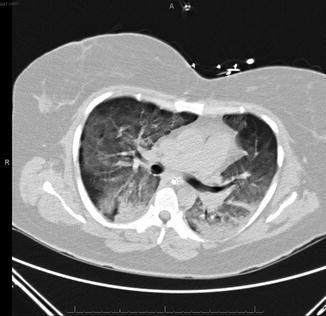
Fig. 21.2
Representative section of chest CT from patient in case study demonstrating bilateral alveolar infiltrates with mild compressive atelectasis in the dependent lung zones
Question
What approach should guide this patient’s ventilator management?
Answer
Lung Protective Ventilation
All patients with the acute respiratory distress syndrome should be treated with lung protective ventilation in order to avoid ventilator associated lung injury (VALI). This patient was started on assist control mechanical ventilation with a tidal volume of 350 ml and 100 % FiO2. Neuromuscular blockade with cisatracurium was initiated. The depth of paralysis was monitored with train of four nerve stimulation and the depth of sedation with midazolam and fentanyl was assessed via bi-spectral analysis. Over the next 12 h her PEEP was increased to 16 cm H2O and her FiO2 was decreased to 40 %. During this time the patients plateau airway pressure ranged between 26 and 28 cm H2O. She was treated with broad spectrum antibiotics, vancomycin, pip-tazo and azithromycin. Results of a culture obtained from a mini-BAL specimen failed to grow any pathogenic organisms. Cisatracurium was discontinued after 48 h. At that time, solu-medrol was begun at a dose of 1 mg/kg body weight. The patient had had already been on an insulin drip but the glucose target range was changed to less than 110 mg/dL from the usual less than 150 mg/dL at that time. Daily sedation holidays were instituted to assess mental functioning and a physical therapy consult was initiated to promote mobility. The patient remained hemodynamically stable with good renal function and diuresis with furosemide was initiated, resulting in a negative fluid balance of 2400 ml on the third ICU day and about 1–2 L/day subsequently. Gas exchange remained satisfactory such that on the fourth ICU day PEEP was decreased to 5 cm H2O. By that time the patient was able to march in place at the bedside and take a brief walk into the hall outside of her room. Later that day the patient was weaned and extubated. She was transferred out of the ICU to the general medical ward the following day.
Principles of Management
Risk Factors for ARDS and Diagnosis
Several risk factors for the development of ARDS have been identified. The lung injury prediction score (LIPS) is a model which incorporates know risk factors and predicts the likelihood of developing ARDS accordingly [1] (Table 21.1). Many patients with multiple risk factors do not develop ARDS as even patients with a LIPS score of more than 7 only develop ARDS less than half of the time. In the case presentation above the patient’s history of chronic alcohol ingestion was a predisposing risk factor for the development of ARDS in what was likely to have been an aspiration pneumonia, which itself in another risk factor.
Table 21.1
Lung injury prediction score (LIPS)
LIPS points | Examples | |
|---|---|---|
Predisposing conditions | (1) Patient with history of alcohol abuse with septic shock from pneumonia requiring Fi O2 > 0.35 in the emergency room: Sepsis + shock + pneumonia + alcohol abuse + Fi O2 > 0.35 1 + 2 + 1.5 + 1 + 2 = 7.5 | |
Shock | 2 | |
Aspiration | 2 | |
Sepsis | 1 | |
Pneumonia | 1.5 | |
High-risk surgerya | ||
Orthopedic spine | 1 | |
Acute abdomen | 2 | |
Cardiac | 2.5 | |
Aortic vascular | 3.5 | |
High-risk trauma | (2) Motor vehicle accident with traumatic brain injury, lung contusion, and shock requiring Fi O2 > 0.35 Traumatic brain injury + lung contusion + shock + Fi O2 > 0.35 2 + 1.5 + 2 + 2 = 7.5 | |
Traumatic brain injury | 2 | |
Smoke inhalation | 2 | |
Near drowning | 2 | |
Lung contusion | 1.5 | |
Multiple fractures | 1.5 | |
Risk modifiers | ||
Alcohol abuse | 1 | |
Obesity (BMI > 30) | 1 | (3) Patient with history of diabetes mellitus and urosepsis with shock Sepsis + shock + diabetes 1 + 2–1 = 2 |
Hypoalbuminemia | 1 | |
Chemotherapy | 1 | |
Fi O2 > 0.35 (>4 L/min) | 2 | |
Tachypnea (RR > 30) | 1.5 | |
SpO2 < 95 % | 1 | |
Acidosis (pH < 7.35) | 1.5 | |
Diabetes mellitusb | −1 |
The PaO2 to FiO2 (P/F) ratio, a measure of oxygenation impairment was part of the old American European Consensus Conference diagnostic criteria for acute lung injury and ARDS. Although easily calculated the P/F ratio did not account for the effect of mean airway pressure on oxygenation. The Berlin criteria for ARDS, published in 2012 [2], did away with the concept of acute lung injury (ALI) in favor of classifying ARDS as mild, moderate or severe. ARDS severity is based on oxygenation criteria which also accounts to some extent, for the application of positive airway pressure. The diagnosis of ARDS is based on clinical presentation and physiology. Diffuse alveolar damage is usually seen histopathologically, but may be absent even in cases of severe ARDS [3].
Berlin Definition of ARDS
Timing
Within 1 week of a known clinical insult or new or worsening respiratory symptoms
Chest Imaging
Bilateral opacities – not fully explained by effusions, lobar/lung collapse, or nodules
Origin of Edema
Respiratory failure not fully explained by cardiac failure or fluid overload
Need objective assessment (e.g. echocardiography) to exclude hydrostatic edema if no risk factor present
Oxygenation
Mild ARDS – The PaO2/FiO2 is >200 mmHg, but ≤300 mmHg, on ventilator settings that include positive end-expiratory pressure (PEEP) or continuous positive airway pressure (CPAP) ≥5 cm H2O.
Moderate ARDS – The PaO2/FiO2 is >100 mmHg, but ≤200 mmHg, on ventilator settings that include PEEP ≥5 cm H2O.
Severe ARDS – The PaO2/FiO2 is ≤100 mmHg on ventilators setting that include PEEP ≥5 cm H2O
Lung Protective Ventilation
Avoiding over distension of the lung during mechanical ventilation of ARDS patients is called lung protective ventilation (LPV). LPV reduces hospital and 28 day mortality [4], presumably by decreasing lung inflammation and avoiding the fibrinoproliferative phase of this condition. The largest trial to date demonstrating this was done by the NIH sponsored ARDS Network, whose results were published in 2000 [5]. The approach undertaken in this trial, the utilization of a tidal volume of less than 6.5 cc/kg of ideal body weight (IBW) (but at least 4 cc/kg) and maintaining a plateau pressure of less than 30 cm of H2O has become the standard treatment to provide mechanical ventilation to patients with ARDS. The calculation of ideal body weight is based on height:
Plateau airway pressure is not a threshold variable [6] and it should be maintained as low as realistically possible, even though in the ARDSNet trial tidal volume was allowed to increase up to 8 cc/kg as long as plateau pressure remained under 30 cm H2O. The ARDS Network approach is the standard approach to lung protective ventilation. However, multiple other approaches have been championed, each ostensibly offering a refinement of the basic lung protective ventilation approach of the ARDSNet. In a retrospective analysis of several of the large trials, a low driving pressure (ΔP) was found to be better correlated with ARDS mortality than tidal volume or plateau pressure [7]. Driving pressure is the pressure being applied by the ventilator to distribute gas to the recruited portion of the lungs which are not collapsed from compressive atelectasis caused by the weight of the lung in the dependent lung zones. In a patient not making spontaneous respiratory efforts ΔP can be estimated as plateau pressure minus PEEP in cm H2O.
Open Lung Ventilation
Avoiding alveolar overdistension with a low tidal volume is the established mechanism of avoiding ventilator-associated lung injury. In the supine ARDS patient delivered gas is distributed to non-dependent lung zones. Dependent lung zones are not ventilated due to the weight of the lung, i.e. compressive atelectasis. In between the two zones is an area of lung which distends and collapses with each delivered ventilator breath, a phenomenon termed cyclic atelectasis. In animal models cyclic atelectasis produces lung injury. Optimizing the recruitment of additional areas of collapsed lung with PEEP in order to mitigate the effect of cyclic atelectasis is the rationale behind open lung ventilation. Meta-analysis of several large clinical trials demonstrated a mortality benefit to open lung ventilation [8]. However, each of the three large trials used for this analysis individually failed to demonstrate a mortality benefit. To the extent that the large clinical trials did not demonstrate harm with higher levels of PEEP, such as an increased rate of pneumothorax, clinicians may choose to use the open lung approach in their patients. Several approaches to performing open lung ventilation are available. A study evaluating the effect of PEEP on lung recruitment, as evaluated by CT scan, suggested the best tradeoff between lung recruitment while simultaneously avoiding lung overdistension was obtained via a high PEEP strategy similar to that utilized in the Lung Open Ventilation Study (LOVS) [9] (Table 21.2).
Table 21.2
Example of open-lung high positive end-expiratory pressure (PEEP) strategy
FIO2 | 0.3 | 0.4 | 0.5 | 0.6 | 0.7 | 0.8 | 0.9 | 1.0 |
PEEP | 5–10 | 10–18 | 18–20 | 20 | 20 | 20–22 | 22 | 22–24 |
Prone Ventilation
A large randomized, multi-center trial, PROSEVA, demonstrated an impressive mortality benefit to patients who underwent prone ventilation 18 h per day [10]. Prone ventilation can be considered to be a form of open lung ventilation. This trial addressed criticisms of earlier, negative trials of prone ventilation in that the study subjects with ARDS had a severe oxygenation defect, were proned for long periods of time daily and a protocolized lung protective approach was used in the control group, the original ARDS network approach [11]. Using the low PEEP ARDS Network approach in the control group, while appropriate, leaves open the question as to whether proning adds incremental benefit to patients who are already receiving higher levels of PEEP (see open lung ventilation, below). In addition, patients underwent neuromuscular blockade, which itself may have a beneficial effect on outcome. Despite these concerns, prone ventilation has assumed an important place as a rescue modality in the treatment of severe ARDS patients who have not responded to a conventional lung protective strategy.
Related posts:


Full access? Get Clinical Tree


