 Used to obtain cerebrospinal fluid (CSF) and to measure the opening pressure of the subarachnoid space to aid in the evaluation and management of patients with acute headache or other symptoms of the following conditions:
 Meningitis
 Subarachnoid hemorrhage (SAH)
 Carcinomatous meningitis
 Pseudotumor cerebri (occasionally for spontaneous intracranial hypotension)
 Occasionally for Guillain–Barré syndrome, multiple sclerosis, inflammatory demyelinating polyneuropathy
 Occasionally in cases of encephalitis
CONTRAINDICATIONS
 Patients who need a lumbar puncture (LP) and have any of the following should first have a brain imaging study, indicating that it is safe to perform an LP:
 Altered mental status
 Papilledema
 New focal neurologic examination abnormalities
 Elevated intracranial pressure (ICP)
 Age 60 years or older (relative)
 Immunocompromised
 Seizure within 1 week before presentation
 Exception, pseudotumor cerebri (where by definition, the ICP is elevated)
 Suspicion of spinal cord mass, or epidural hematoma/abscess
 Skin or soft-tissue infection overlying lumbar spine
 Anatomic abnormalities: For example, patients with lumbar hardware from prior spinal surgery
 Coagulopathic patients
RISKS/CONSENT ISSUES
 Post-LP headache occurs in approximately 15% to 20% of patients. Using an atraumatic or noncutting spinal needle decreases the incidence of this, and multiple therapies exist to treat this specific type of headache.
 The procedure can cause local pain. Local anesthesia will be given.
 Needle puncture can cause local bleeding, which is usually minimal
 Potential for introducing infection exists; however, this is extremely rare. Sterile technique will be utilized.
 Theoretical risk of damage to neural tissue exists. Such occurrences are also very rare, most often temporary, and affect spinal nerve roots, not the cord itself.
LANDMARKS
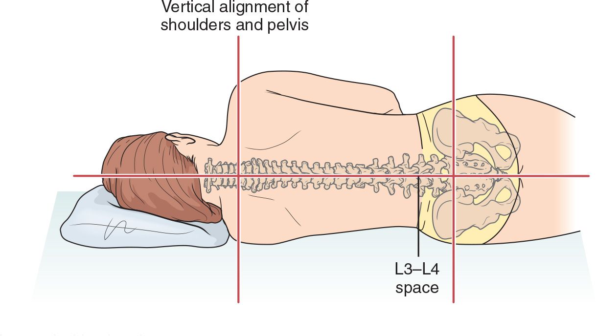
The transverse axis connecting iliac crests passes through L4 vertebral body, allowing for identification of the L4-5 and L3-4 interspaces (FIGURE 45.1)
TECHNIQUE
 Patient Preparation
 Explain the procedure to the patient and obtain patient consent
 Position the patient in either lateral decubitus or sitting position (the sitting position may be easier to use but precludes accurate ICP measurement)
 Lateral Decubitus Position
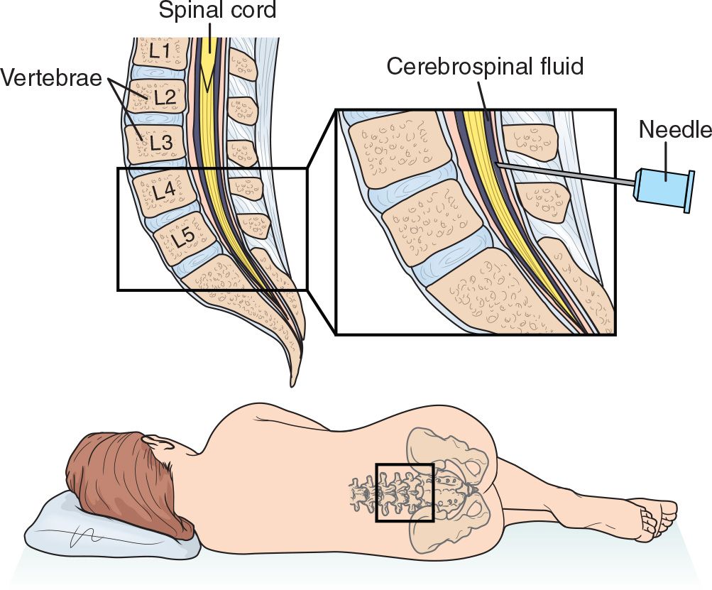
 Have the patient lie on one side, with knees to chest and head/shoulders curled toward knees as much as possible. Placing a pillow under the head helps reduce twisting of the shoulders.
 Ensure that the lumbar spine lies parallel to the edge of the bed. (In an infant/child, or a poorly cooperative adult, it will be necessary to have an assistant hold the patient securely in the optimal position.) The top shoulder and hip should be directly above their bottom counterparts.
 A cooperative patient can be asked to curve his/her lower back, out like an “angry cat,” to optimally open the spinous processes
 Sitting Position
 Have the patient sit on the side of the bed with the bed positioned below patient’s midthigh and with the feet of the patient touching floor, if possible
 Ask the patient to curve the torso forward over a bedside table positioned in front of him/her; table height should be level with patient’s upper abdomen. A pillow may be placed on the table for patient’s comfort.
 After positioning, but before prepping, mark the target for needle insertion with firm pressure from the Luer-lock (hub) end of a needle sheath (or some other blunt device such as a pen) firmly against the skin (which will leave a mark for several minutes and provides a visual target)
 Prepare a wide area with povidone–iodine or chlorhexidine gluconate solution
 Ensure the sterile field includes L4-5 and L3-4 interspaces. (Should first attempt at L4- 5 be unsuccessful, the L3-4 interspace will be readily accessible.)
 Use sterile drapes to frame workspace
 Reassess landmarks. It is crucial that the midline be defined.
 Sometimes, in overweight patients, feeling the spinous processes in the thoracic spine (where they are easier to palpate) and marching down will help the clinician ensure that they are in the midline
 Ultrasonography has been shown to reduce the failure rate, number of attempts, and traumatic punctures and can be used as an adjunct



Full access? Get Clinical Tree


