Fig. 42.1
Computer telemetry to access the status of the pump before pump refill
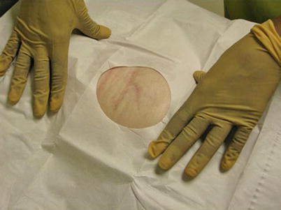
Fig. 42.2
After carefully prepping the abdomen on multiple occasions, sterile drapes are placed
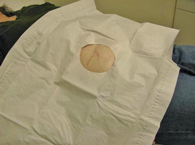
Fig. 42.3
The patient should be assessed to orientation of the pump, and as to evidence of any skin abnormalities
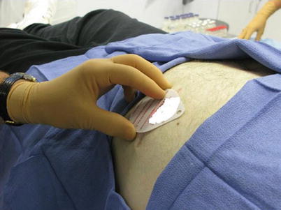
Fig. 42.4
In some cases, laser-guided fluoroscopy can be used to refill the pump. This may be very helpful in the obese patient or in a patient with an abnormal abdominal wall secondary to scar or poor tissue integrity
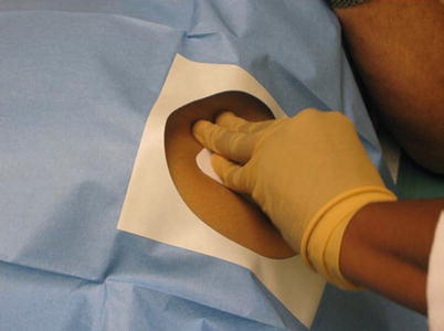
Fig. 42.5
Sterile dressings are placed once the pump is refilled
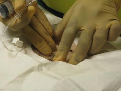
Fig. 42.6
A template can be used to help identify the port of the intrathecal pump
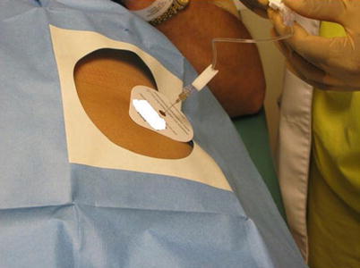
Fig. 42.7


After securing the needle in the lumen, the pump should be aspirated and compared to the expected volume. One should hold the needle in place
Related posts:


Full access? Get Clinical Tree
