Key Clinical Questions
What are the fundamental goals of cardiopulmonary resuscitation?
Which components of resuscitation are considered vital to success?
How can the common pitfalls of resuscitation be surmounted?
What treatments should be instituted immediately upon successful resuscitation?
How should outcomes in resuscitation shape the discussion of advanced directives?
Introduction
This chapter focuses on cardiopulmonary arrest and the events immediately antecedent to this life-threatening event. Cardiopulmonary resuscitation is a time-dependent, team-based effort to reverse physiologic events that may culminate in a patient’s imminent death. Biblical and ancient Egyptian hieroglyphic texts allude to mouth-to-mouth ventilation in divine contexts, but other texts indicate Jewish midwives used mouth-to-mouth resuscitation as early as 3300 years ago to revive stillborn children.
Since standardization of closed chest cardiac massage was first described systematically in the medical literature in 1960, chest compressions have remained the only reliable means of reviving a patient in cardiopulmonary collapse.
In the United States an estimated 375,000 to 750,000 hospitalized patients suffer in-hospital cardiac arrest (IHCA) requiring advanced cardiac life support (ACLS) annually. The incidence of IHCA is estimated to be as high as 1–2% of all patients admitted to academic hospitals with a prevalence of approximately 65 people per 100,000 nationally.
IHCA encompasses a spectrum of disorders, from insufficient cardiac output to generate appreciable cerebral perfusion, including arrhythmia or shock, to complete cessation of cardiac activity. Vital sign anomalies can often herald impending inpatient cardiac arrest by minutes to hours, but many cardiac arrests occur suddenly and without warning. Acute pulmonary arrest may precede IHCA and can occur iatrogenically related to sedative or opiate analgesics overdose.
Triage
Since standardization of closed chest cardiac massage (CCCM) was first described systematically in the medical literature in 1960, CCCM has remained the only reliable means of reviving a patient in cardiopulmonary collapse. It is an effective and powerful intervention that, when unnecessarily delayed, can lead to poor patient outcomes. In one study, survival dropped from 34% to 14% if CCCM was delayed even as little as 60–120 seconds from the time the patient collapsed. Therefore, clinicians must recognize and respond to cardiac arrest immediately for resuscitation measures to be effective.
Advanced cardiac life support (ACLS) combines basic life support (BLS) measures with specific interventions such as medication, defibrillation, transthoracic pacing, and advanced airway management. This chapter focuses on (1) the techniques that are essential to successful cardiopulmonary resuscitation, neurologic recovery (as defined by the cerebral performance category 0–1), and (2) decision making based on patient resuscitation status.
While often considered adequate for credentialing purposes, completion of American Heart Association courses fails to result in long-term meaningful skill performance. Health care providers’ capabilities to demonstrate appropriate technique for CCCM and capabilities to successfully navigate the steps of cardiopulmonary resuscitation begin to degrade just weeks following course completion. Therefore, for the whole medical team to respond concisely and in a coordinated fashion, clinicians must have extensive medical knowledge, training, drilling practice, continued education, and feedback.
Many providers are reluctant to initiate CCCM without complete assurance that the patient is truly in cardiopulmonary arrest (confirmed by vital signs or electrocardiographic rhythm), often leading to unnecessary delays in initiation of potentially life-saving treatment. Furthermore, fundamental pulse assessment, even in nonemergency situations, is not consistently accurately evaluated by clinicians. One study tasked providers to determine whether or not patients had palpable pulses during elective cardiopulmonary bypass surgery. Ultimately providers took around 20 seconds to assess the pulse and were less than 70% accurate.
Time spent gathering cardiac monitoring, attaching leads, and setting up equipment can further delay promptly needed interventions to prevent death. In fact, clinicians may need to initiate CCCM prior to confirming cardiopulmonary arrest. Prompt initiation of CCCM for any patient who appears to be in extremis (ie, unarousable, or clinically unstable with suspicion of cardiopulmonary arrest) should occur until confirmatory evaluation, often by a multispecialty resuscitation team, offers a high degree of confidence that CCCM can be discontinued. Providers should share a culture of support that accentuates that the greater harm to patients is in failing to initiate CCCM in contrast to the potential harms of CCCM (rib fracture, pneumothorax, organ perforation, etc).
|
Pathophysiology
Cardiopulmonary arrest heralds death and may be an expected outcome in many hospitalized patients. However, rarely is cardiopulmonary arrest the first manifestation of physiologic events that ultimately culminate in collapse: patients frequently have alteration in mental status or significant vital sign changes (pyrexia, hypotension, bradycardia, decrease in oxygen saturation, change in respiratory rate, etc), often hours before developing cardiac arrest. Intervention during this pre-arrest period may prevent cardiac arrest altogether. Alternatively, health care personnel may identify patients who are at the end of life and may thus benefit from a meaningful discussion about limiting resuscitative measures, including offering “Do Not Resuscitate” or “Allow Natural Death” orders. Many patients are not well informed about the resuscitative process and may have inflated images of routine successful resuscitation shaped from popular culture embodied by television and film. Clinicians often perform cardiopulmonary resuscitation on patients without informed consent—a discussion of the relevant risks, benefits, and alternatives to therapy along with the clinicians’ recommendations. Thus the pre-arrest period may offer an unparalleled opportunity to give patients an active role in deciding whether resuscitation is desired.
While no one specific condition results in cardiopulmonary collapse, many health care–associated interventions predispose patients to arrest and often require minimal intervention early on to alter the course of catastrophe (Table 121-1). Intervention during impending cardiac arrest requires a detailed history of recent interventions ranging from invasive procedures to recent sedation or anesthesia.
Cause | Intervention |
|---|---|
Hypoxia due to medication or anesthesia | Supportive oxygen, reversal agents (naloxone for opiates, flumazenil for benzodiazepines) |
Acidemia due to hypercapnic respiratory failure from medication or obstructive sleep apnea | Ventilation support (noninvasive or mechanical ventilation) |
Pulmonary embolism | Appropriate VTE prophylaxis (pharmacologic unless significant contraindication); high index of suspicion and timely treatment |
Cardiac arrhythmia due to acute coronary syndrome | Appropriate early intervention including antiplatelet therapy, beta-blockers, anticoagulation and early percutaneous coronary intervention (PCI) |
Hyperkalemia | Calcium, sodium bicarbonate, insulin with dextrose, consideration of early hemodialysis; check for acid–base derangements |
Hypokalemia | Correction of magnesium (first) followed by potassium; check for acid–base derangements |
QT prolongation | Attention to medications known to prolong the QT interval (such as fluoroquinolones) and consideration of cardiac monitoring |
Hypotension from severe sepsis | Early massive volume resuscitation with consideration of inotropes |
Anticipated end-of-life care | Discussion of appropriate “Do Not Resuscitate” or “Allow Natural Death” orders and palliative care in appropriate patients |
Responses to inpatient emergencies require multiple individuals who take on individual roles and integrate as a team. For care to function effectively and seamlessly during health care emergencies, each clinician must assume a narrowly focused essential function or task (such as assessing a patient’s airway, recording data in a flowsheet, or ensuring chest compressions are adequate) and perform the task with high quality to facilitate the best possible patient outcome engendered by the team as a whole.
Recognizing that early intervention in impending cardiopulmonary arrest may prevent the arrest altogether, many hospitals have implemented rapid response teams (RRTs), consisting of any combination of critical care nurses, respiratory therapists, pharmacists, and/or physicians to attend to patients who have some parameters of clinical instability but are not yet in extremis. While data remain mixed about the success of RRTs to stave off cardiopulmonary arrest, most data suggest RRTs facilitate earlier discussion of patient advanced directives and improve communication with critical care teams and intensivists, potentially reducing mortality and need for crisis activation of cardiopulmonary arrest teams (ie, code teams). Speculation exists that RRTs have not yet realize their full outcome potential because some staff still hesitate to call an RRT when indications to do so are present.
|
Computerized medical records with “involuntary” initiation of an RRT due to deviations in certain predefined criteria (heart rate or blood pressure instability, depression of mental status, or decrease in oxygen saturation) may allow RRTs to fulfill their full potential and may reduce code team activation and improve patient outcomes. In the interim, hospitalists must foster a culture of safety where any provider can initiate an RRT for any reason without fear of reprisal or judgment. Hospitalists should always thank other clinicians for calling RRTs and keeping the patients’ safety of the utmost concern.
Respiratory arrest from medications (anesthesia, benzodiazepines, or opiates) can lead to cardiac arrest through hypoxia and changes in the pH due to combined metabolic and respiratory acidosis. Respiratory arrest is often masked for some time due to the ubiquity of oxygen administration in hospitalized patients, which can lead to a prolonged period of hemoglobin oxygenation while ventilation may have already stopped. Overreliance on pulse oximetry as a sole source of interpreting ventilation effort may delay response to respiratory arrest until the patient is hypoxic and profoundly acidemic. Systemic hypoxia causes pulmonary artery constriction, right ventricular failure, and systemic hypotension from poor right heart output coupled with loss of vascular tone from hypoxia (circulatory shock).
Gasping is a powerful agonal respiration that improves patient survival through increased cardiac output, improved oxygenation, improved ventilation, and improved cerebral blood flow. Positive pressure bag-valve-mask (BVM) ventilations may confer a negative effect on the ability of patients to gasp suggesting a role for delaying aggressive airway management until later on in a cardiac resuscitation. Conversely, in pure respiratory arrest, early aggressive airway management to prevent deterioration into cardiac arrest may be warranted.
|
Cardiac arrest can occur from multiple distinct mechanisms. True cardiac arrest (cardiac standstill) occurs either as a primary mechanism (from arrhythmias like ventricular fibrillation that prevent normal cardiac function) or as a secondary mechanism (from asystole or from an extended period of failed resuscitation and cardiac myocyte death). Most cardiopulmonary arrest episodes do not occur due to true cardiac standstill but rather from marked impairment in cardiac output resulting in systemic arterial hypotension, tissue hypoxia, and organ failure. Precardiac, intracardiac, or postcardiac mechanisms may independently or in combination result in cardiopulmonary arrest (Table 121-2).
Precardiac | Intracardiac/intrapulmonary | Postcardiac |
|---|---|---|
|
|
|
Once appropriate resuscitation equipment has arrived, clinicians can begin to differentiate whether the cardiac arrest is due to a “shockable” or “nonshockable” cardiac rhythm.
Transthoracic electrical shocks can terminate some pathological cardiac rhythms that inhibit normal cardiac function. These can include ventricular fibrillation, ventricular tachycardia, AV nodal reentrant tachycardia, atrial fibrillation, and atrial flutter. While ventricular fibrillation has a very characteristic pattern, the other rhythms may be difficult to differentiate during an emergency and in the absence of 12-lead electrocardiography. In the setting of an unconscious patient in extremis, all of these rhythms are considered pathologic and warrant immediate electrical shock.
Despite recommendations by the International Liaison Committee of Resuscitation (ILCOR) (the subsection of the American Heart Association responsible for publication of the ACLS guidelines) that differentiation of the exact cardiac arrhythmia may dictate very different types of cardiac intervention, ranging from dose (in joules) of electrical therapy to medication selection, confirming an exact rhythm diagnosis may not be practical. Thus it is reasonable to treat all of these rhythms similarly in a cardiopulmonary arrest in the event of clinical uncertainty. Fundamentally similar to administration of CCCM, delays in electrical therapy can significantly negatively impact patient outcomes with even minimal delays. If a patient is not critically ill, then time allows for conscientious assessment of cardiac rhythm via 12-lead electrocardiogram (ECG) with appropriately targeted therapies for the underlying arrhythmia (see Chapters 126 and 128 on supraventricular tachyarrhythmias and ventricular arrythmias).
|
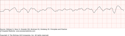
Ventricular fibrillation results from disorganized myocardial electrical activity, and the heart is unable to generate a contraction to produce cardiac output. Hospitalists should be able to identify ventricular fibrillation confidently on rhythm strip (Figure 121-1).
The characteristic physiologic phases of ventricular fibrillation arrest underscore the importance of rapid electrical therapy. During the first few minutes of ventricular fibrillation (reflecting the combination of the “acute” and “electrical” phases of arrest, lasting up to 5–6 minutes), the myocardium is highly responsive to counter shock. This explains in part why successful defibrillation is so common on commercial airlines and in casinos where employees are trained to rapidly attach and initiate automated external defibrillators (AEDs). The acute and electrical phases can be extended when CCCM is initiated promptly, thus underscoring how critical CCCM is as an immediate therapy while definitive defibrillation equipment is located, attached, and initiated.
In the absence of CCCM, patients will degenerate into the “circulatory” phase where electrical therapies are less effective due to progressive tissue hypoxia and myocyte death. During this phase, CCCM must be performed for several minutes antecedent to attempts at defibrillation.
Unchecked, patients will eventually enter the “metabolic” phase of ventricular fibrillation starting around the tenth minute of cardiac arrest. While there remains a slim hope of successful cardiac resuscitation at this point, in the absence of effective CCCM irreversible brain damage occurs and survival to hospital discharge rapidly becomes improbable.
Ventricular tachycardia resulting in cardiopulmonary arrest fundamentally is identical to ventricular fibrillation in treatment: CCCM and early electrical shock are indicated.
Perhaps the most overwhelming change to resuscitation in recent years is the acknowledgment of severe ventricular stunning following electrical shock. For several minutes following defibrillation—and extending for a variable duration thereafter—the heart is mechanically dysfunctional and unable to generate an adequate cardiac output for organ perfusion or brain function. Consequently, it is absolutely critical to reinitiate CCCM for 1–2 -minutes after defibrillation whether or not the shock is successful at aborting the ventricular arrhythmia.
|
Related posts:
 Strategies for Cost-Effective Care
 Building, Growing, and Managing a Hospitalist Practice
 Designing a Hospitalist Compensation and Bonus Plan
The Face of Health Care Emerging Issues for Hospitalists
Designing a Hospitalist Compensation and Bonus Plan
The Face of Health Care Emerging Issues for Hospitalists
 Medical Malpractice
Preventing and Managing Adverse Patient Events: Patient Safety and the Hospitalist
Medical Malpractice
Preventing and Managing Adverse Patient Events: Patient Safety and the Hospitalist

Full access? Get Clinical Tree


