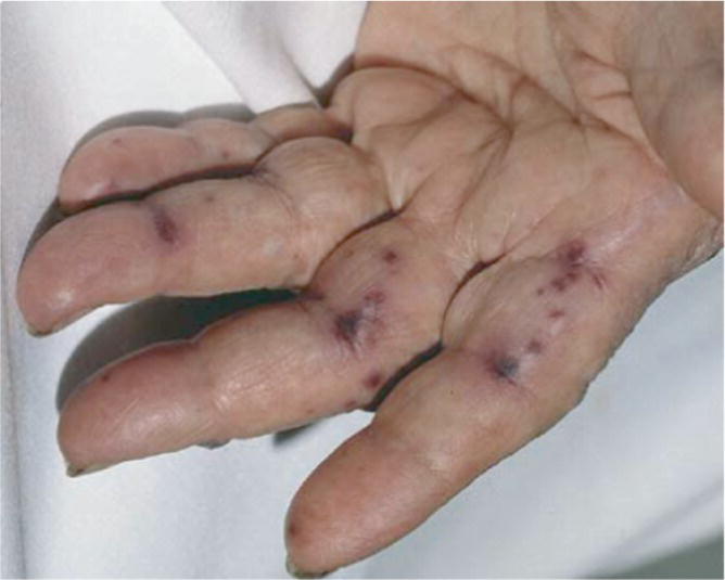
W. Mason Bonner and Jonathan Sheele Department of Emergency Medicine, Mayo Clinic, Jacksonville, FL, USA Infective endocarditis is a bacterial infection of the endothelial lining of the heart which begins as an infection of cardiac valve leaflets and can evolve to include other portions of the heart and vasculature. These vegetations can cause devastating illness when they form abscesses or embolize to extracardiac sites. In industrialized countries, the incidence is ~3–9 cases per 100,000 population with a male to female predilection of roughly 2:1. Classically, endocarditis has been associated with intravenous drug abuse and patients with underlying cardiac valvular pathology (rheumatic heart disease, congenital cyanotic valvular disease). Mortality associated with endocarditis is high with some estimates approaching 18%.1 Healthcare‐associated illness is increasingly recognized as a cause and accounts for roughly one‐fourth to one‐third of cases. Patients with prosthetic valves, indwelling catheters, and intracardiac devices represent a growing percentage of endocarditis cases. The most common complications of endocarditis include heart failure, stroke, nonstroke embolization, and intracardiac abscess. The mitral and aortic valves are more commonly affected and account for >70% of cases. Left‐sided endocarditis overall tends to carry a worse prognosis than right‐sided disease. The most common presenting symptom is fever which is present in up to 80% of cases. Relapsing fever >5 days without a clear source increases the possibility of endocarditis. Other presenting findings include new murmur (48%), worsening known murmur (20%), hematuria (26%), and splenomegaly (11%).1 More uncommon but well‐known cutaneous and mucous membrane manifestations of disease such as splinter hemorrhages, Janeway’s lesions (Figure 26.1), and Roth spots occur in fewer than 10% of patients. Other presentations can include sepsis, meningitis, new heart failure, and symptoms of embolization such as pulmonary embolus (PE), stroke, peripheral arterial occlusion, hepatic abscess, and renal failure.3 The most common pathogen causing endocarditis is Staphylococcus and this is followed by Streptococcus.4 Together they account for 80% of cases of infectious endocarditis. Gram‐negative bacteria are less frequently implicated, and total around 5% of cases and can involve Haemophilus, Aggregatibacter, Cardiobacterium, Eikenella, and Kingella, referred to as HACEK.5 Fungal infections represent 1–2% of cases. Emergency providers must maintain a high index of suspicion for endocarditis because most cases are not confirmed in the emergency department. Figure 26.1 Janeway’s lesions from bacterial endocarditis. (Fitzpatrick et al. [2], Reproduced with permission from McGraw‐Hill Education.) What is the most accurate method to diagnose suspected infective endocarditis in the ED? Diagnosis of endocarditis in the ED relies primarily upon effective patient risk stratification and the most validated tool is the modified Duke criteria.6 The Duke criteria have a sensitivity and specificity approaching 80% and have a strong negative predictive value.7 Initially proposed in 1994 by Durack et al., the Duke criteria aided clinicians in their approach to the diagnosis of endocarditis.8
Chapter 26
Infective Endocarditis
Background
Clinical question


Full access? Get Clinical Tree


