 Indicated only if location of foreign body (FB) is certain
 Removal may be done in 30 minutes or less
RELATIVE CONTRAINDICATIONS
 Involvement of joint—orthopedic consultation may be required
 Coagulopathies or bleeding diathesis
 Allergy to anesthetic
 Chronic medical problems that delay healing, such as diabetes, uremia, or immunocompromised state
 Involvement of abdomen/pelvis/thorax
 Near major vascular structures that are difficult to visualize
 Uncooperative, difficult, or intoxicated patient
 FB not localized
RISKS/CONSENT ISSUES
 Procedure can cause pain (local anesthesia will be given)
 Local bleeding
 There is potential for introducing infection (sterile technique will be utilized)
 Risk of injuring local neurovascular structures
 Scar at site of FB removal
 Patient must be informed that all FBs may not be removed
 Retained wood FBs always develop an inflammatory response, but retained bullets rarely produce inflammation
 General Basic Steps
 Localize the FB
 Patient preparation
 Decide on method of removal
TECHNIQUE
 Localize the FB
 Get multiple projections of plain x-ray using a soft-tissue technique (e.g., underpenetrated film); to locate radiopaque FBs, place a marker (i.e., needle) on the skin surface at the wound entrance before the x-ray procedure
 Although glass and metal are easily located with plain films, ultrasonographic localization may be required for wood and thorns
 All intraorbital and intracranial FBs must be imaged by computed tomography (CT)
 If a patient has a previously explored wound demonstrating signs of infection, poor wound healing, or persistent pain, consider doing a CT
 Patient Preparation
 Sterilize and drape the area from where FB will be removed
 Anesthetize area either via local infiltration or appropriate nerve block
 General Removal Techniques
 Enlarge the entrance to wound with an adequate skin incision
 Spread the soft tissue with hemostats, avoiding use of fingers
 Hemostats can help find glass in a wound by creating a clicking sound when tapped against glass
 If visualization is inadequate, consider excision of small block of tissue, only if no significant neurovascular structures are involved
 When searching for a thorn or needle, consider an elliptical incision, undermine the skin in all directions, and then compress the sides, expelling the FB
 Closure of the wound after thorough irrigation is indicated unless exploring a contaminated wound
TICK REMOVAL
 Nonmechanical means of tick removal is not recommended (i.e., drowning the tick in petroleum jelly), because it may cause the tick to regurgitate, increasing infection risk (FIGURE 84.1)
 Mechanical removal
 Using the tip of forceps, grab the tick as close as possible to the patient’s skin, and apply steady traction
 Ensure that all mouth parts are removed. Use an 18-gauge needle to remove retained pieces.
 Thoroughly cleanse the area with soap and water
 In patients at high risk of Lyme disease, consider administration of 200 mg of doxycycline in a single dose (or amoxicillin in pediatrics) (Figure 84.1)
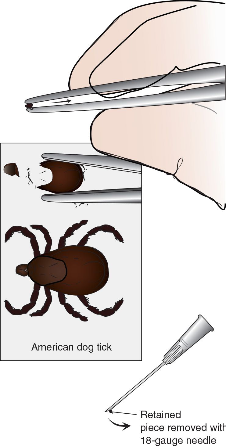
FIGURE 84.1 Tick removal. (From Bond GR. Envenomation management and tick removal. In: Henretig FM, King C, eds. Textbook of Pediatric Emergency Procedures. Philadelphia, PA: Williams & Wilkins; 1997:1328, with permission.)



Full access? Get Clinical Tree


