Nasal bleeding that does not stop with a topical vasoconstrictor and direct pressure applied by the patient (described below in the “Technique” section).
PROCEDURES
 Cauterization
 Anterior source bleeding that can be visualized
 Anterior Nasal Packing
 Anterior source bleeding that cannot be visualized
 Cauterization fails
 Posterior Nasal Packing or Balloon Tamponade
 Anterior source cannot be identified and
 Bleeding from both nares or
 Blood draining into the posterior pharynx
 Anterior packing of both nares fails to control bleeding
CONTRAINDICATIONS
 None
None
RISKS
 Nasal packing can cause pain or discomfort
 Risk of infection, septal damage, and ulceration
 Risk of balloon migration, airway obstruction, or aspiration with posterior packing
LANDMARKS
 The choice of procedure depends on the source of bleeding. The source can be identified by direct visualization, or with use of a nasal speculum (described in more detail in the “Technique” section).
 Nosebleeds are most commonly anterior (80% to 90%), originating from the Kiesselbach plexus in the septum
 Most posterior nosebleeds originate from branches of the sphenopalatine artery in the posterior nasal cavity or nasopharynx. A posterior source is likely when an anterior source cannot be identified, when bleeding is from both nares, or when blood is draining into the posterior pharynx.
 General Basic Steps
 Patient preparation
 Analgesia
 Visualization
 Cauterization
 Anterior packing
 Posterior packing
TECHNIQUE
 Patient Preparation
 Ask the patient to blow his or her nose, which decreases the effects of local fibrinolysis and removes clots (FIGURE 75.1)
 Ask the patient to lean forward and apply continuous pressure to the alae of both nares for 10 to 20 minutes. If this stops bleeding, consider sending patient home with follow-up to ear, nose, and throat (ENT) specialist or Primary Medical Doctor (PMD).
 Analgesia
 Apply a topical anesthetic and vasoconstrictor
 Soak cotton or gauze in 2% lidocaine with or without topical epinephrine or 4% topical cocaine. Place in the nasal cavity for 15 to 20 minutes.
 Alternatively, a topical anesthetic with a decongestant (2% lidocaine and 4% phenylephrine mixed 1:1) or oxymetazoline hydrochloride (Afrin) nasal spray may be used
 Because patients may be apprehensive and packing is uncomfortable, opiates or benzodiazepines may be given before the examination (strongly recommended in cases of posterior packing)
 Visualization
 Position the patient sitting upright, facing forward in the sniffing position
 Insert a nasal speculum so that one blade moves superiorly and the other inferiorly and spread the nares vertically. Suction all remaining clots and blood. This permits visualization of most anterior sources.
 Cauterization
 If an anterior source is visualized, cauterization should be attempted. To be effective, cautery should be performed after bleeding is controlled. Only cauterize one side of the septum at a time to prevent perforation or necrosis of the septum.
 Chemical cauterization can be performed using silver nitrate sticks. Apply the tip of a silver nitrate stick to the bleeding site until a white precipitate forms (usually a few seconds, rarely more than 10).
 If bleeding is vigorous, electrocauterization can be used. Apply the device to the bleeding site for up to 10 seconds or until a white precipitate forms.
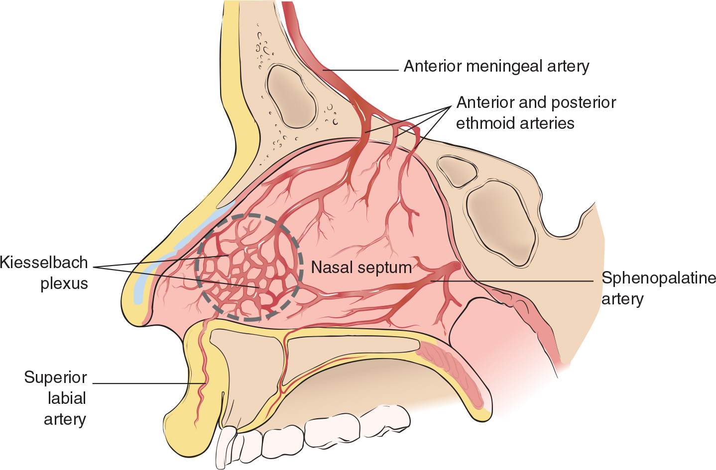
FIGURE 75.1 Anatomy of nasal septum. (From Kost SI, Post JC. Management of epistaxis. In: Henretig FM, King C, eds. Textbook of Pediatric Emergency Procedures. Philadelphia, PA: Williams & Wilkins; 1997:663, with permission.)



Full access? Get Clinical Tree


