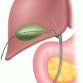
Fig. 2.1
Three different types of percutaneously placed endoscopic enteral tubes: (a) typical PEG with inner and outer flange, single port, (b) PEG with jejunal tube extension, double port for gastric aspiration, distal jejunal feeding, (c) direct percutaneously placed endoscopic jejunostomy
Administer prophylactic broad spectrum antibiotics prior to the procedure.
Decompress the stomach of contents prior to decompressive enteral tube placement to minimize aspiration risk.
Potential Pitfalls
Aspiration during an endoscopic procedure.
Visceral Injury with percutaneous endoscopic technique.
Bleeding along the newly established enteral access tract.
Operative Strategy
Prior to providing long-term enteral access, the surgeon must have a clear understanding of whether pre-pyloric or post-pyloric feeding is appropriate. In most instances, a percutaneous endoscopic approach with good technique can provide safe long-term enteral access to meet a patient’s needs with morbidity and discomfort less than that associated with open or even laparoscopic surgery. Nevertheless, alternative surgical techniques are sometimes needed. In certain cases, it may even be appropriate to have multiple different plans available for a single operative encounter to provide a feeding tube for a given patient, e.g., PEG (Plan A), possible laparoscopic assisted PEG or a laparoscopic G-tube (Plan B), or even an open G-tube placement (Plan C). It is therefore crucial to be familiar with a number of different techniques as well as different types of feeding tubes. It is also of paramount importance that appropriate feeding tubes and equipment are available for these procedures and the facility’s staff is familiar with their use and maintenance prior to performing a given procedure.
In general, the only two conditions that will preclude a percutaneous endoscopic approach to providing long-term enteral access include the inability to access the gastrointestinal tract with an endoscope (e.g., obstructing oropharyngeal mass or trauma, esophageal obstructing tumor) and the inability to establish a safe window for percutaneous tube placement. In this context, a safe window is a site where the feeding tube can be passed percutaneously through subcutaneous tissues, abdominal wall musculature, and gastric or jejunal wall with minimal chance of inadvertently damaging adjacent visceral structures. In these two situations—no endoscopic access or no safe window, a laparoscopic approach to feeding tube placement can generally be utilized as a viable alternative procedure.
Operative Technique
Endoscopic Techniques
PEG
The pull technique for PEG placement can be performed using a one operator and one nurse/technician assistant approach. The patient should be positioned supine with supplemental oxygen and monitoring devices in place. A diagram of patient, operator, and assistant positioning is presented (Fig. 2.2). The abdomen should be generously prepped and draped as the safe window of PEG placement is not always strictly in the left upper quadrant—it can be epigastric and rarely even just to right of the patient’s midline. The PEG kit and associated equipment should then be opened and ready. Many commercial vendors provide different versions of these kits and not every kit will have everything needed. If the kit is anticipated to provide for everything including an endoscopic snare, scissors, etc., then this must be checked and ensured prior to starting the procedure. After the appropriate administration of conscious sedation or monitored anesthesia care, the operator introduces a flexible upper endoscope into the esophagus and a brief, standard EGD examination is performed. After ensuring no unexpected pathology, insufflation of the gastric body is provided sufficient to efface the rugal folds.


Fig. 2.2
A surgeon using this setup can perform both the endoscopic component and abdominal placement component of the procedure while an assistant (RN or GI Technician) assists by activating the transillumination feature and running the endoscopic snare
At this point, the crux of the procedure is finding the safe window for tube placement. There are three evaluations which should routinely be conducted to ensure such a safe window is found prior to PEG placement.
1.
The operator can use the back of the plunger of a syringe of anesthetic (e.g., 1 % Lidocaine) to carefully palpate with this sterile instrument thereby finding a site with good 1:1 ratio of palpation that is discrete in appearance (Fig. 2.3).


Fig. 2.3
The surgeon/endoscopist palpates the abdominal wall using the back of the plunger so as to maintain sterility of the field even as she runs the endoscope. This demonstrates discrete, 1:1 palpation at site of potential PEG placement, aiding in establishing a safe window for percutaneous placement
2.
The endoscope is then driven up to approach the greater curve, placing the tip of the scope on the anterior gastric wall. This will then achieve transillumination, casting a warm/orange glow across the abdominal wall seen at the level of skin at the site of anticipated PEG placement. In morbidly obese patients, the assistant may need to press/deploy the transilluminate button so as to provide sufficient intensity of lighting to transilluminate a truly long abdominal wall distance. This button/feature is not generally needed in patients of normal body habitus.
3.
The needle of the syringe of anesthetic is then passed into the lumen of the stomach at the site of 1:1 palpation and transillumination. While passing this needle through the subcutaneous, abdominal wall, and visceral tissues, the syringe is aspirated to ensure no bubbles are found until the needle enters the hollow gastric lumen visualized endoscopically. If no bubbles are seen until the needle enters the stomach lumen, this is deemed a “safe track” for PEG tube placement. If bubbles or gas is aspirated prior to entry into the stomach, this track is not safe and either another track should be found or the PEG procedure potentially aborted (Fig. 2.4). In performing the safe track aspiration, it is very helpful to ensure that the operator watches the screen to ensure passage of the syringe into the gastric lumen even as the assistant watches the syringe to notify the team when bubbles are first observed within the syringe.”


Fig. 2.4
(a) This demonstrates a negative safe track aspiration with the anesthetic/sounding needle, helping to assure a safe window for percutaneous placement of a PEG even as (b) demonstrates a positive safe track aspiration suggesting viscera between the stomach and abdominal wall, potentially despite apparent 1:1 palpation
Authorities have traditionally used the lack of transillumination during this procedure as a contraindication for completing the PEG procedure as planned. If however there is good 1:1 palpation and the safe track is negative for any bubbles prior to entry into the gastric lumen at the site, there are series which have demonstrated the safety of completing the procedure as planned in this setting. Thus at least two of these three criteria must be found to establish the safe window for PEG placement.
An endoscopic snare is then passed through the endoscope and is left waiting in the gastric lumen. With this done, a 1 cm incision is made through skin at the selected site. A catheter is passed through the incision along the same trajectory as the 1:1 palpation and aspiration anesthetic needle into the stomach lumen. The snare is used to encircle the catheter thus securing it. The operator passes the looped wire through the catheter into the gastric lumen, taking care to orient it properly. The snare is then moved from where it is secured on the catheter to the wire. Once the snare is securely around the wire, the entire endoscope along with snare and wire are removed from the patient’s mouth. The wire is then freed from the endoscope channel and secured to the tip of the PEG tube. The wire is then pulled (thus PULL technique) at the level of the abdominal wall, bringing the wire and the now secured tip of the PEG tube completely out of the patient until gentle traction or resistance is felt as the inner flange abuts the gastric wall (Fig. 2.5). Once positioned, an outer flange is placed down the tube (typically resting at 2–4 cm on the skin of a normal body habitus patient), the wire and some of the excess length of tubing is cut free and the cap is applied. At this point, a repeat endoscopic evaluation may be performed to ensure the inner flange is sitting without undue tension and no bleeding is visualized.


Fig. 2.5


(a) This sagittally depicts the endoscopic snaring of the transabdominal, trans-gastric wire which has been passed through a catheter. This wire, snare, and endoscope are then pulled back out of the oropharynx. (b) This depicts the pulling of the wire, now secured to the PEG tube, back out and across the abdominal wall until the inner flange is felt to abut the gastric and abdominal wall

Full access? Get Clinical Tree


