 Penetrating Chest Trauma
 Traumatic arrest with witnessed signs of life* in the field
 Persistent hypotension (systolic blood pressure (SBP) <60 mm Hg) despite resuscitative efforts
 Blunt Trauma
 Traumatic arrest that occurs in the emergency department (ED)
 Persistent hypotension (SBP <60 mm Hg) despite resuscitative efforts
 Pulmonary Trauma
 Chest tube drainage >1,500 mL
 Persistent hypotension or cardiac arrest with known lung laceration
 Air Embolism
 Persistent signs of hypovolemic shock
 Hemoptysis and cardiac arrest after intubation and ventilation
 Nontraumatic Hypothermic Cardiac Arrest
 In settings where cardiopulmonary bypass is not immediately available
 Goals
Goals
 Relief of cardiac tamponade
 Support of cardiac function with open massage, cross-clamping the aorta, and/or internal cardiac defibrillation
 Control of hemorrhage
 Diagnosis and management of air embolism
 Mediastinal irrigation and rewarming (for hypothermic cardiac arrest)
CONTRAINDICATIONS
 No signs of life and prehospital cardiopulmonary resuscitation (CPR) performed:
 >15 minutes after penetrating trauma
 >10 minutes after blunt trauma
 Multisystem blunt trauma
 Severe head injury
 Asystole as an initial rhythm without tamponade
 Inability to provide definitive care after procedure
RISKS/CONSENT ISSUES
 This is an emergent procedure and does not require written consent
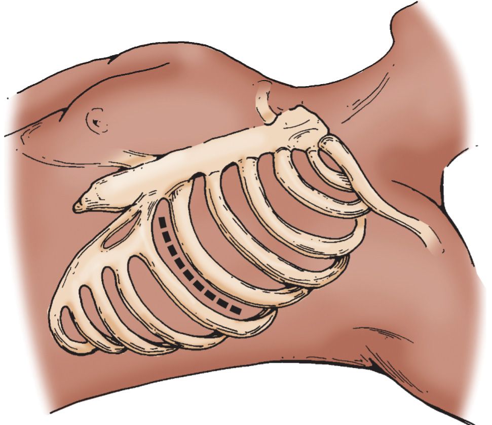
LANDMARKS (FIGURE 15.1)
 Left-sided supine anterolateral approach over the 5th rib, in the fourth intercostal space
 In males incise below the nipple
 In females below the inframammary fold
 General Basic Steps
 Incision
 Dissection and rib spreading
 Pericardotomy
 Cardiac massage
 Hemorrhage control
 Aortic cross-clamping
TECHNIQUE
 Patient Preparation
 Patient should be intubated and a nasogastric tube should be placed (this should not delay the procedure!)
 Place towels under the left chest and place left arm above the head
 Sterilize the incision area with copious povidone–iodine solution
 Incision
 Using a no. 20 blade, incise from the sternal border to the posterior axillary line
 During the primary incision, cut firmly through subcutaneous tissue to the intercostal muscle
 Dissection and Rib Spreading
 Using scissors, cut the intercostal muscles above the 5th rib to avoid the neurovascular bundle
 Temporarily stop ventilation just before exposing the pleura to avoid lacerating the lung
 Insert rib spreader with the ratchet placed toward the axilla and handlebar down
 Use a Gigli saw, Lebsche knife, or trauma shears to cut the sternum for right-sided exposure
 Pericardiotomy
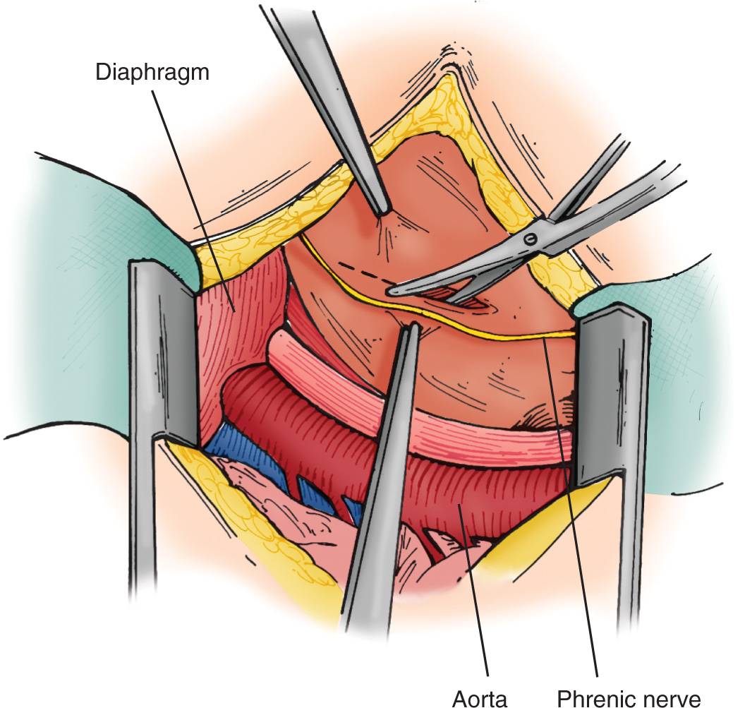
 Hold the pericardium with forceps, and use scissors to cut from the cardiac apex to the aortic root (FIGURE 15.2)
 The incision should be made anterior and lateral, avoiding the left phrenic nerve
 Evacuate blood and clots from the pericardium
 Deliver the heart from the pericardium if cardiac repair is required



Full access? Get Clinical Tree


