 Clinical suspicion of acute anterior, posterior, lateral, medial, or divergent dislocation with or without neurovascular compromise
 The clinical presentation depends on the type of dislocation
 Suspected dislocation is clinically confirmed by disruption of the relationship between the tip of the olecranon and the distal epicondyles of the humerus in comparison with the unaffected elbow
 Radiographic evidence of anterior, posterior, lateral, medial, or divergent dislocation (FIGURE 65.1)
CONTRAINDICATIONS
 Open dislocations require emergent consultations with an orthopedic surgeon
 Multiple failed reduction attempts with adequate sedation should prompt consultation with an orthopedic surgeon
 Irreducible elbow dislocations may require operative management
 An elbow that has been unreduced for 7 or more days will likely require open reduction with an orthopedic surgeon
RISKS/CONSENT ISSUES
 Procedural sedation may be associated with loss of airway reflexes and respiratory arrest (these risks are extremely rare)
 Soft-tissue injury may occur with reduction attempts
 Fractures and neurovascular injury may occur with reduction attempts
 General Basic Steps
 Obtain necessary x-rays
 Sedation/Analgesia
 Position patient
 Reduction
 Postprocedure exam/x-rays
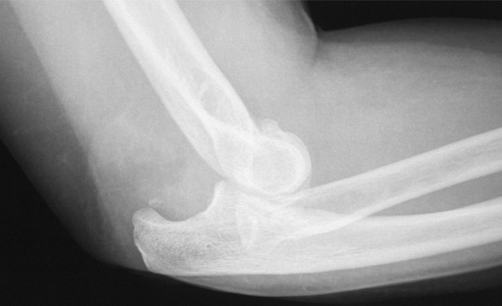
FIGURE 65.1 Posterior dislocation of the olecranon. (From Campbell C. Elbow dislocation. In: Greenberg MI, ed. Greenberg’s Text-Atlas of Emergency Medicine. Philadelphia, PA: Lippincott Williams & Wilkins; 2005:492, with permission.)
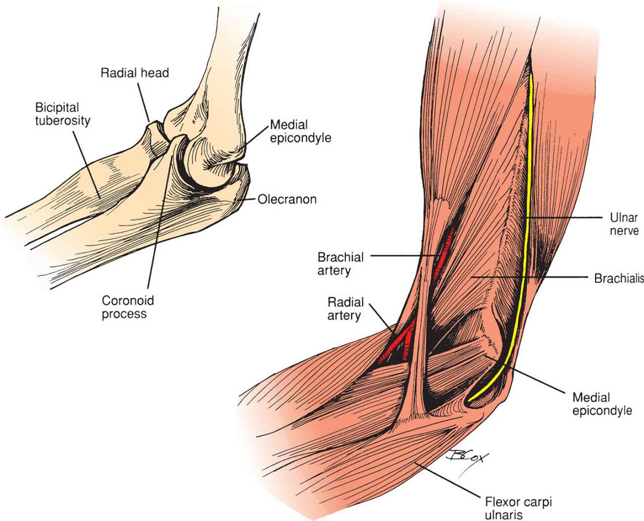
FIGURE 65.2 Elbow anatomy. (From McCue FC III, Sweeney T, Urch S. The elbow, wrist, and hand. In: Perrin DH, ed. The Injured Athlete. 3rd ed. Philadelphia, PA: Lippincott Williams & Wilkins; 1999, with permission.)
TECHNIQUE
 Perform a complete neurovascular check before any reduction attempt
 Obtain radiographs of the affected joint and consider radiographs of one joint above and below the injury (shoulder and wrist)
 Complex dislocations (those with associated fractures) may require consultation with orthopedic surgery
 Dislocations with neurovascular compromise should be reduced without prior imaging
 Anesthesia/analgesia: Consider parenteral analgesics. Reduction may also be attempted with injection of local anesthetic alone into the elbow joint or an ultrasound-guided brachial plexus block
 Reduction technique is determined by the type of dislocation
TECHNIQUE: POSTERIOR DISLOCATION
 80% to 90% of all elbow dislocations
 Mechanism of injury: Most commonly caused by a fall on an outstretched hand with the arm in extension
 Clinical presentation: Shortened forearm that is held in flexion with a prominent olecranon posteriorly. In addition, a defect may be palpable above the olecranon (FIGURE 65.2).
 Associated injuries:
 Fractures including radial head and coronoid process are common
 Small fractures of the coronoid process may be treated as simple posterior dislocations
 Neurologic symptoms accompany 15% to 22% of dislocations
 Ulnar nerve injury is most common followed by median nerve injury
 Radial nerve injury commonly occurs when the dislocation is complicated by radial head fracture
 Traction leading to stretch injury, local swelling, and entrapment during reduction are common causes of nerve injury
 Brachial artery injury occurs in 5% to 13% of posterior dislocations
 Reduction Techniques
 Supine Technique
 Place patient in supine position
 An assistant stabilizes the humerus by wrapping both hands around arm just distal to axilla
 The physician grasps the wrist with one hand and places the other hand just above the antecubital fossa with the thumb on the olecranon (FIGURE 65.3)
 The physician applies slow, steady in-line traction while the assistant applies steady countertraction
 To minimize additional trauma to the coronoid process, the elbow is held in slight flexion and the wrist is held in supination as traction is applied
 Avoid hyperextension as this may cause injury to the median nerve or brachial artery
 Reduction is accompanied by a “clunk” that is heard or felt
 Alternatively, the forearm may be gently flexed in an effort to reduce the joint
 Seated Technique
 Patient is seated in a high backed chair with arm hanging over the back of the chair in a flexed position
 The physician applies traction by gently pulling down on the patient’s hand while guiding the olecranon into place using the other hand
 The physician may also elect to simply apply downward pressure onto the olecranon to reduce the elbow
 Reduction is once again signaled by a “clunk”
 This method has the advantage of requiring only a single physician
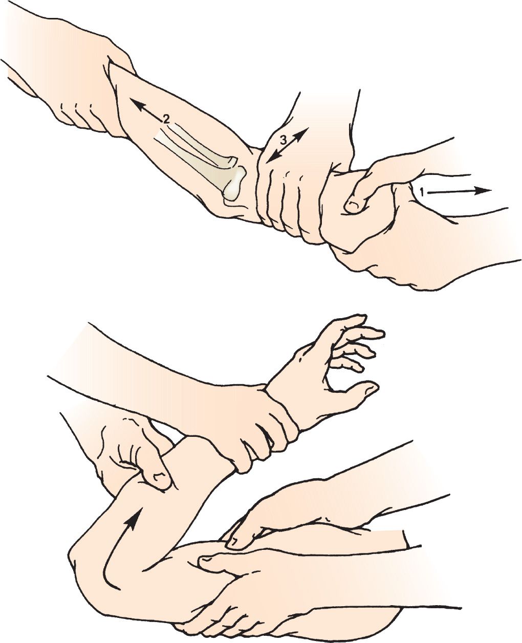
FIGURE 65.3 Technique for reduction of posterior dislocation of the elbow. (From Perron AD, Germann CA. Elbow injuries. In: Wolfson AB. Harwood-Nuss’ Clinical Practice of Emergency Medicine. 6th ed. Philadelphia, PA: Lippincott Williams & Wilkins; 2015:260, with permission.)



Full access? Get Clinical Tree


