 Clinical suspicion of joint dislocation
 Incidence of dislocations: Dorsal proximal interphalangeal (PIP) >> volar PIP >> dorsal metacarpal (MCP) thumb > dorsal MCP finger >> volar MCP dorsal distal interphalangeal (DIP) >> volar DIP
 Radiographic evidence of dislocation
CONTRAINDICATIONS
 Complex dislocation—rupture of or entrapment in the joint of ligaments or tendons surrounding the joint requires open reduction and repair
 Chronic dislocation (>3 weeks duration)
 Open dislocation
 Unstable joint
 Multiple failed reduction attempts can convert a simple dislocation into a complex dislocation and prompt urgent orthopedic consult
 General Basic Steps
 Neurovascular examination
 Prereduction radiograph
 Analgesia
 Reduction
 Postreduction neurovascular examination
 Immobilization
 Postreduction radiograph
LANDMARKS
 Nerves run on the lateral surface of each digit at the 2, 4, 8, and 10 o’clock positions
 Flexor tendons run on the volar surface of the digit
 Extensor tendons run on the dorsal surface of the digit
 PIP joint
 Dorsal dislocation: The fibrous volar plate resists dorsal dislocations (FIGURE 68.1)
 Complex dislocation: Head of proximal phalanx or ruptured volar plane can become entrapped in the joint space (FIGURE 68.2)
 Volar dislocation: Three bands of the extensor tendon (central slip, radial and ulnar lateral bands) resist volar dislocations (FIGURE 68.3)
 Complex dislocation: Extensor slip tendons can rupture and become entrapped within the joint space (FIGURE 68.4)
 Lateral dislocation: Radial collateral ligaments resist ulnar dislocation, ulnar collateral ligaments resist radial dislocation
 DIP and thumb IP: Mechanisms of dislocation and reduction are anatomically analogous
RISKS/CONSENT ISSUES
 Risks in joint reduction include swelling with permanent joint enlargement, residual pain, stiffness, or deformity and conversion into complex dislocation including fracture
 Risks in anesthesia
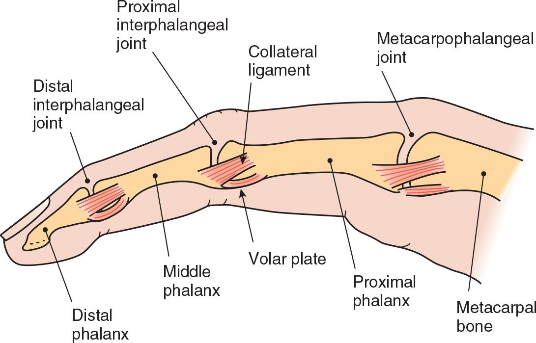
FIGURE 68.1 Fibrous volar plates are found at the MCP and IP joints where they reinforce the joint capsules and limit hyperextension. (From Leggit, JC, Meko CJ. Acute finger injuries: Part I. Tendons and ligaments. Am Fam Physician. 2006;73(5):810–816.)
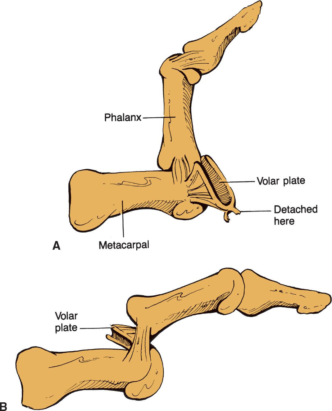
FIGURE 68.2 Complex dorsal PIP dislocation indicated by (A) rupture of PIP volar plate and (B) entrapment of volar plate fibers and head of proximal phalanx into the joint space. (Jackimczyk K, Shepherd SM, Blackburn P. Hand injuries. In: Wolfson AB, ed. Harwood-Nuss’ Clinical Practice of Emergency Medicine. 5th ed. Philadelphia, PA: Williams & Wilkins; 2009, with permission.)
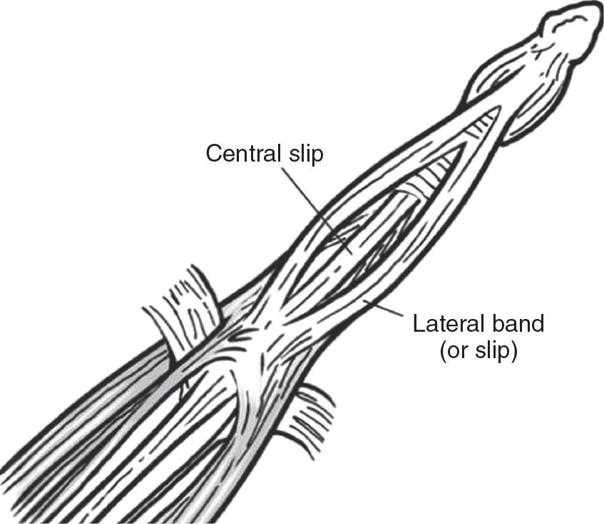
FIGURE 68.3 Extensor tendon mechanism trifurcates at the dorsum of the proximal phalanx (Reichman EF. Emergency Medicine Procedures. 2013. Permission pending.)
SUPPLIES
 Anesthesia
 Reduction: 3″ Webril or gauze padding, 3″ plaster roll, 3″ Ace bandage, aluminum finger splint, adhesive tape, scissors
NEUROVASCULAR EXAMINATION
Any deficit indicates a complex dislocation and urgent orthopedic consultation
 Inspection
 Exclude open dislocation
 Complex dislocation presents with less angulated deformities, with skin dimpling, or rotational deformity of the involved phalanges
 Palpation—frank fracture fragments
 Sensation—normal two-point discrimination on finger pad is between 2 to 4 mm
 Vascular—radial and ulnar pulse-Doppler signal
 Range of motion with stress—assess joint stability with the finger in full extension and in moderate flexion. If displacement occurs, the joint is unstable.
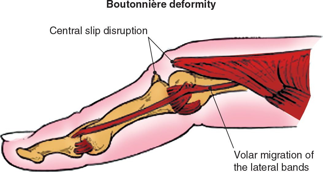
FIGURE 68.4 Central slip of the extensor tendon can rupture in sudden IP hyperflexion and can require operative repair. (From Leggit JC, Meko CJ. Acute finger injuries: Part I. Tendons and ligaments. Am Fam Physician. 2006;73(5):810–816.)



Full access? Get Clinical Tree


