A sensitive way to evaluate for intra-abdominal injury in the trauma patient
 In Blunt Trauma
 Unexplained hypotension
 Concern for injury but no obvious indication for laparotomy and serial abdominal examinations are not practical (i.e., unconscious or under anesthesia)
 Equivocal focused abdominal sonography for trauma (FAST) examination and concern for an intra-abdominal injury
 Patient unsuitable for computed tomography (CT) in whom there is concern for intra-abdominal injury
 Concern for mesenteric or hollow viscous injury not seen on CT
 In Penetrating Trauma
 Anterior abdominal stab wound and evidence of fascial penetration in the stable patient with no obvious indication for laparotomy
 To evaluate for hollow organ or diaphragmatic injury in the stable patient
CONTRAINDICATIONS
 Absolute Contraindications
 Indication for an emergent laparotomy
 Relative Contraindications
 Prior abdominal surgery
 Second or third trimester of pregnancy—consider open technique with supraumbilical approach
 Morbid obesity
 Significant ascites
 Coagulopathy
 General Basic Steps
 Prepare patient
 Analgesia
 Technique
 Open
Open
 Incision
 Dissection
 Incise fascia, then peritoneum
 Place dialysis catheter
 Closed
 Needle into abdomen
 Thread guidewire
 Small skin incision
 Thread dialysis catheter
 Aspirate
 Lavage/Drainage of fluid
 Analysis of fluid
LANDMARKS
The incision should be made in the midline, one-third of the way between the umbilicus and pubic symphysis. In the pregnant patient or the patient with a pelvic fracture, the incision should be made in the midline, just above the umbilicus (FIGURE 16.1).
SUPPLIES
 1% Lidocaine with epinephrine, 20 mL with 25-gauge needle, 10-mL syringe
 Sterile towels or drapes, sterile gown and gloves, mask, and eye protection
 Povidone–iodine (Betadine) solution or chlorhexidine
 11-Blade scalpel
 Syringe and needle (for closed technique)
 Flexible guidewire (for closed technique)
 Two clamps and two retractors (for open technique)
 Peritoneal catheter
 1 L warm saline for infusion (for lavage)
 Tubing to let lavage fluid drain
 Suture (for open technique)
TECHNIQUE
 Preparation
 Place a Foley catheter (unless contraindicated)
 Place a nasogastric tube (unless contraindicated) to suction to decompress the stomach
 Gather all instruments and sterile gown/gloves
 Sterilize the abdomen from costal margin to pubis and from flank to flank with povidone–iodine solution (Betadine) or chlorhexidine
 Drape the area with sterile towels or drapes
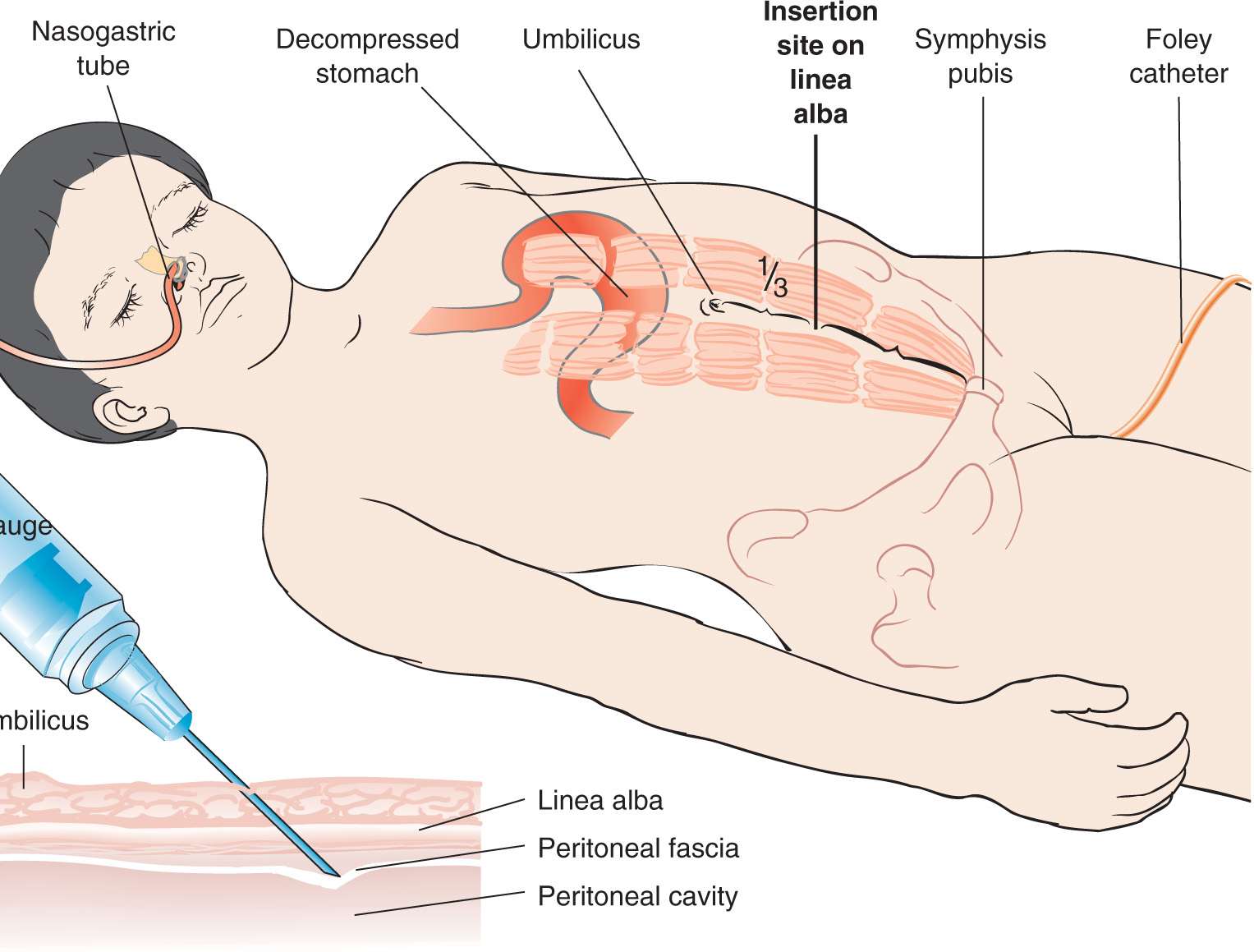
FIGURE 16.1 Anatomical landmarks for diagnostic peritoneal lavage. (From VanDevander PL, Wagner DK. Diagnostic peritoneal lavage. In: Henretig FM, King C, eds. Textbook of Pediatric Emergency Procedures. Philadelphia, PA: Williams & Wilkins; 1997:358, with permission.)



Full access? Get Clinical Tree


