 To provide emergent airway access only when a safer, less invasive airway cannot be established or is contraindicated
 For children younger than 12 years, needle cricothyroidotomy is the surgical airway of choice
CONTRAINDICATIONS
 Absolute
 An oral or nasal airway can be established
 Significant injury or fracture of the cricoid cartilage or larynx (tracheostomy is the procedure of choice)
 Tracheal fracture or transection
 Obstruction below the cricothyroid membrane
 Patients younger than 12 years (needle cricothyroidotomy is the procedure of choice for this age-group)
 Relative
 Neck mass, swelling, or cellulitis
 Neck hematoma
 Coagulopathy
LANDMARKS
 The cricothyroid membrane—an elastic membrane located anteriorly and midline in the neck, measuring 9 mm longitudinally and 30 mm transversely. Bordered superiorly by the thyroid cartilage (“Adam’s apple”) and inferiorly by the cricoid cartilage.
 In children, the larynx is positioned more superiorly than in adults and is relatively smaller in size (FIGURES 6.1 and 6.2)
STANDARD CRICOTHYROIDOTOMY
 Supplies
 Antiseptic solution, drapes, towel clips
 Lidocaine with epinephrine
 No. 11 blade scalpel with handle
 Tracheal hook
 Trousseau dilator
 Tracheostomy tube
 Neck tie or sutures
 Technique
 Preparation
 Hyperextend the neck to more readily identify landmarks, unless the patient has a known or suspected cervical spine injury
 Preoxygenate the patient by bag-mask ventilation
 Test the integrity of the balloon on the tracheostomy tube by injecting with 10 mL of air
 If time permits, apply appropriate antiseptic solution and drape the area with sterile towels
 If time permits and patient is conscious or responding to pain, infiltrate the skin of anterior neck with 1% lidocaine solution with epinephrine
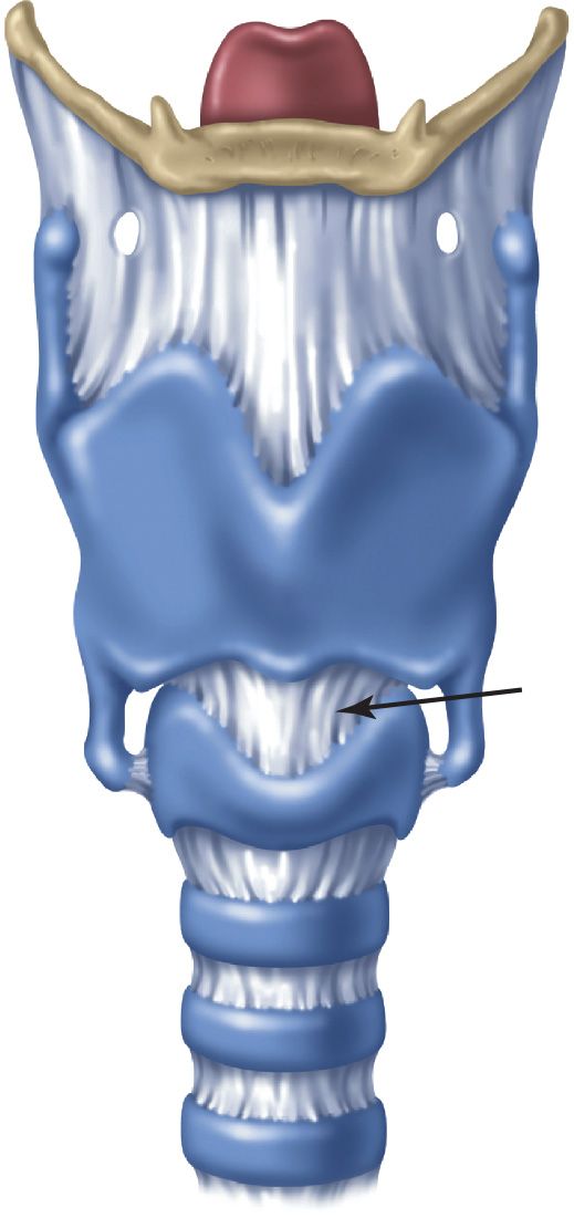
FIGURE 6.1 Anatomy of the larynx. The cricothyroid membrane (arrow) is bordered above by the thyroid cartilage and below by the cricoid cartilage. (From Walls RM, Murphy MF. Manual of Emergency Airway Management. 4th ed. Philadelphia, PA: Lippincott Williams & Wilkins; 2012:162, with permission.)
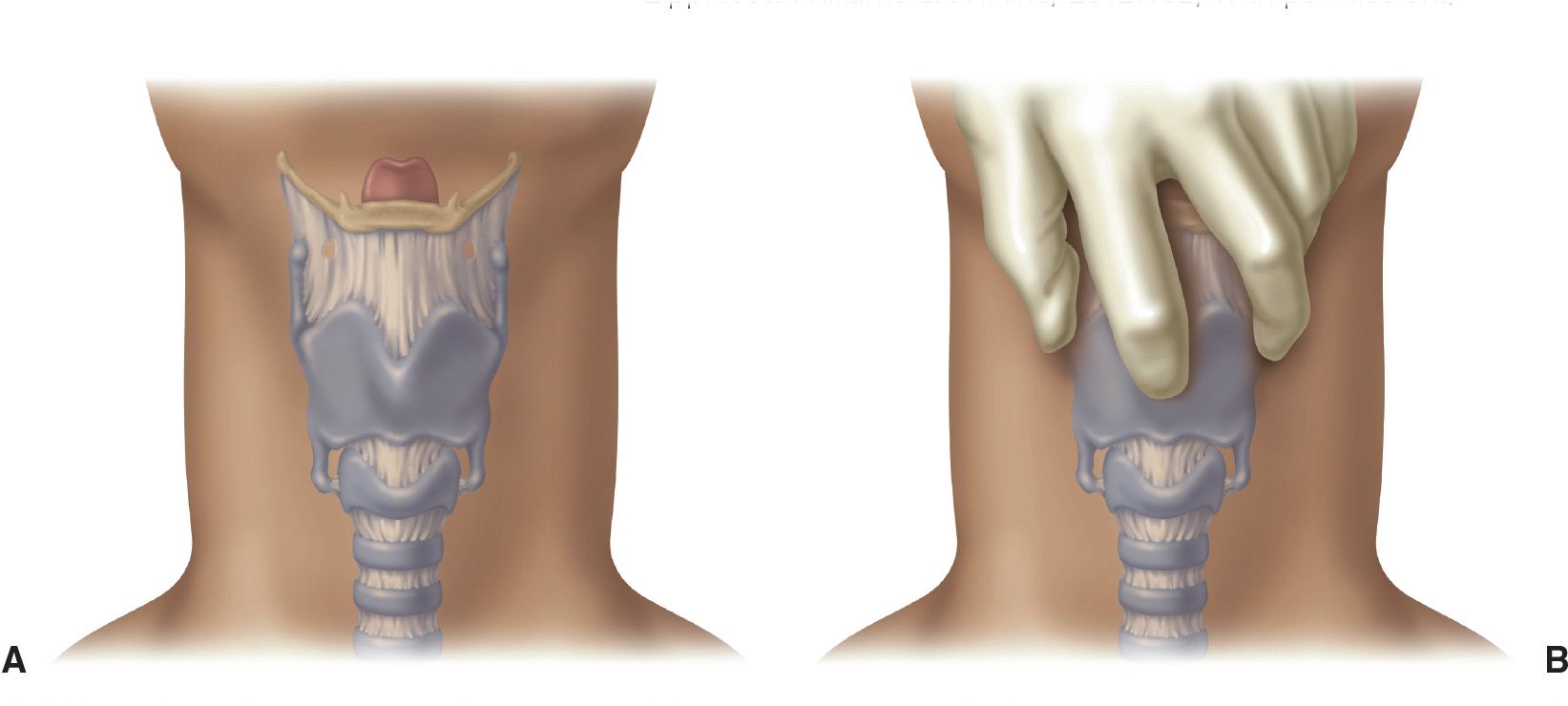
FIGURE 6.2 A: Surface anatomy of the airway. B: The thumb and middle finger immobilize the superior cornua of the larynx; the index finger palpates the cricothyroid membrane. (From Walls RM, Murphy MF. Manual of Emergency Airway Management. 4th ed. Philadelphia, PA: Lippincott Williams & Wilkins; 2012:163, with permission.)
 General Basic Steps
 Identify the landmarks
 Stabilize the larynx
 Incise the skin
 Reidentify the cricothyroid membrane
 Incise the cricothyroid membrane
 Insert the tracheal hook
 Insert the trousseau dilator
 Insert the tracheostomy tube
 Inflate the cuff and confirm tube position
 Secure the tracheostomy tube
 Identify the Landmarks (See Earlier “Landmarks” Section)
 Stabilize the Larynx
 Grasp both sides of the thyroid cartilage with the thumb and middle finger using the nondominant hand
 Palpate the depression over the cricothyroid membrane with the index finger
 Control the larynx throughout the procedure by stabilizing it in this manner and reidentify the cricothyroid membrane at any time during the procedure
 Incise the Skin
 Using a no. 11 scalpel blade in the dominant hand, make a vertical midline incision through the skin and subcutaneous tissue approximately 2 to 3 cm in length
 Care should be taken to extend the incision down to but not through any of the deep structures of the neck
 Reidentify the Cricothyroid Membrane
 Using the index finger of the nondominant hand, reidentify the cricothyroid membrane while maintaining immobilization of the larynx with the thumb and middle finger
 If the cricothyroid membrane cannot be palpated, extend the initial incision superiorly and inferiorly and try to palpate again
 Incise the Cricothyroid Membrane
 Using the stabilizing index finger as a guide, incise the cricothyroid membrane at least 1 cm in length in the horizontal direction
 Note that the skin incision is vertical and the incision through the membrane is horizontal
 Place the index finger into the stoma temporarily while exchanging the scalpel for the tracheal hook
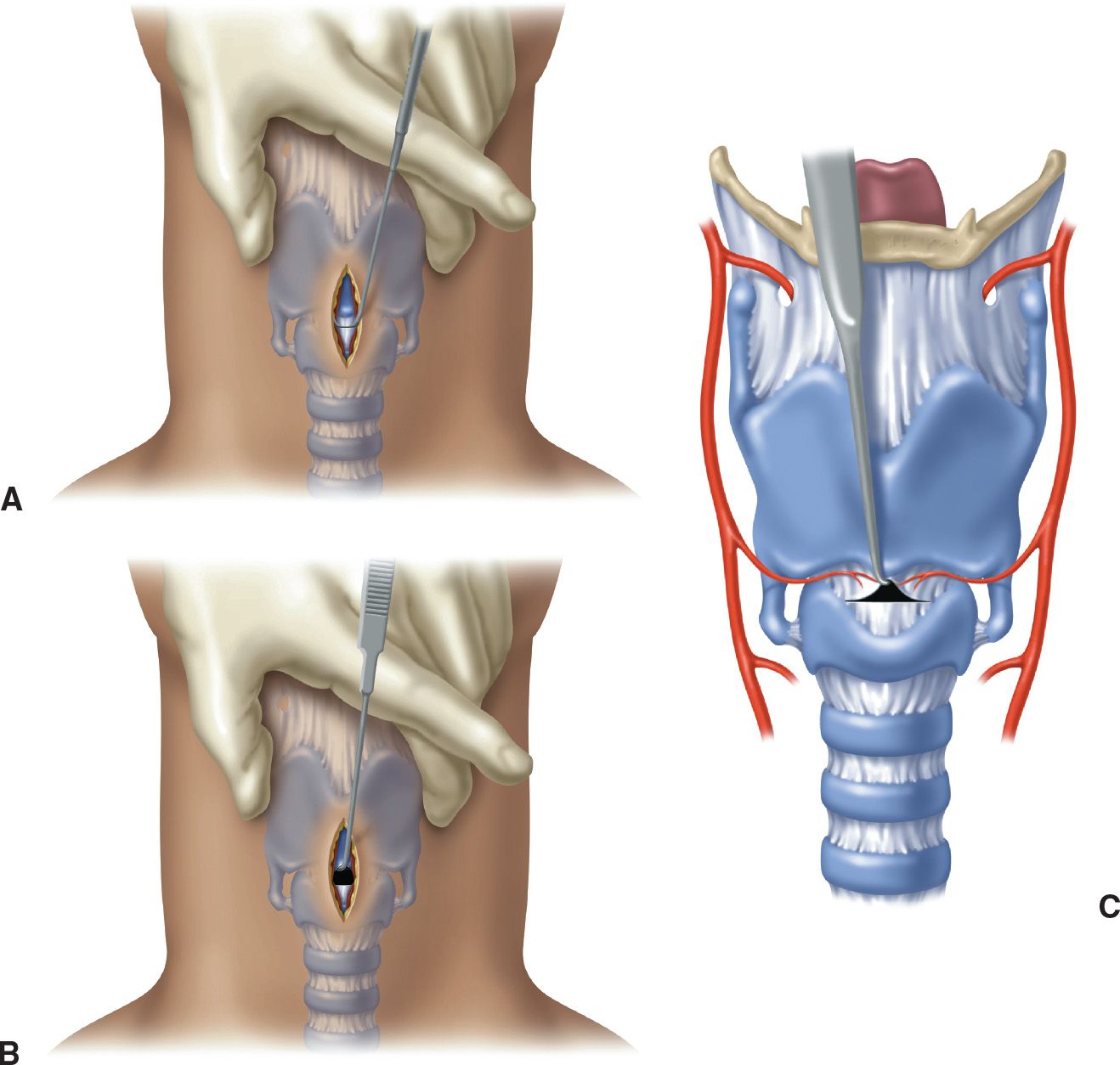
FIGURE 6.3 A: The tracheal hook is oriented transversely during insertion. B and C: After insertion, cephalad traction is applied to the inferior margin of the thyroid cartilage. (From Walls RM, Murphy MF. Manual of Emergency Airway Management. 4th ed. Philadelphia, PA: Lippincott Williams & Wilkins; 2012:166, with permission.)



Full access? Get Clinical Tree


