125 Complications of Gynecologic Procedures, Abortion, and Assisted Reproductive Technology
• Complications with the highest morbidity and mortality are severe hemorrhage, serious infection, damage to intraabdominal structures, and pulmonary embolism.
• Complications seen in the emergency department are usually delayed in presentation, and often difficult to diagnose due to insidious onset, resulting in increased morbidity and mortality and a higher risk for litigation (i.e., ureteral injuries) High suspicion must be maintained.
• Emergency department bedside ultrasonography can provide rapid, early imaging for the evaluation of postprocedural patients, particularly unstable ones.
• Abortion is one of the most common procedures in the United States and overall has very low serious complication rates.
• Ovarian hyperstimulation syndrome is a potentially fatal complication of assisted reproduction in a generally healthy young woman. With no cure, early recognition, aggressive intervention, and close monitoring are key.
This chapter is divided into three main sections—complications of gynecologic procedures, complications following medical and surgical abortion, and complications of assisted reproductive technology (ART).
Gynecologic procedures run the gamut from minor office procedures to major invasive surgery. They can be diagnostic or therapeutic and may initiate pregnancy or terminate it. They represent some of the most common surgical procedures performed in the United States today.
More than 146,000 cycles of ART were reported to the Centers for Disease Control and Prevention from 441 sites in the year 2009. In addition, approximately 600,000 hysterectomies are performed annually, which ranks it behind cesarean section as the most common major surgery in women of reproductive age.2
Complications of Gynecologic Procedures
This section focuses on complications particular to gynecologic procedures that one might encounter in the ED setting and their evaluation and management (Fig. 125.1). Many complications of gynecologic procedures may go unrecognized before discharge, only to be seen later in the ED (Box 125.1). Box 125.2 lists the typical timing of these complications.

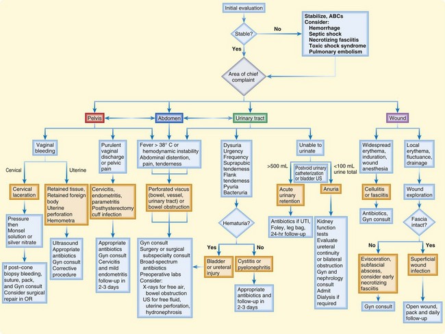
Fig. 125.1 Suggested algorithm for the evaluation and treatment of postoperative gynecologic patients.
ABCs, Airway, breathing, and circulation; Gyn consult, gynecology consultation; OR, operating room; US, ultrasonography; UTI, urinary tract infection.
Box 125.2 Complications of Gynecologic Procedures by Estimated Time Line
Differential Diagnosis and Medical Decision Making
During the evaluation of postoperative patients it is essential to avoid narrowing the differential diagnosis to postoperative complications alone. Other conditions, particularly preexisting ones that may have served as the original indication for surgery (e.g., malignancy, anemia) must be taken into consideration. Laboratory testing and imaging studies should be guided by the differential diagnoses under consideration (see the Priority Actions box).
For patients with complications after a gynecologic procedure, bedside ultrasonography (US) in the hands of a skillful operator can provide rapid recognition of intraabdominal and intrapelvic pathology. Possible ultrasonographic findings include free fluid heralding leakage from a perforated vessel, urinary tract, or viscus (Fig. 125.2); hydronephrosis as a result of ureteral obstruction; a full bladder secondary to urinary retention; fluid collections; and intrauterine contents. US can also be used to guide paracentesis for definitive fluid diagnosis or for the drainage of subcutaneous abscesses. It is important to remember that sensitivity and accuracy are very dependent on the user and interpreter and that anatomy, habitus, and elements such as bowel gas can greatly interfere with adequate imaging. US is a poor modality for evaluating the bowel or retroperitoneal space.

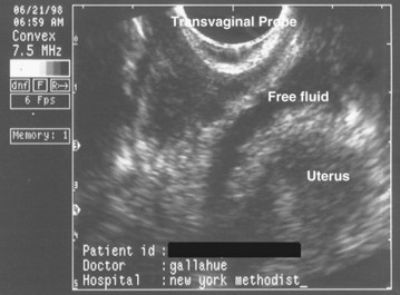
Fig. 125.2 Transvaginal ultrasound image showing free fluid in the cul-de-sac consistent with hemorrhage.
When a patient with postprocedural complications is seen in the ED, the physician who performed the procedure should be contacted; definitive management often requires gynecologic or other surgical intervention.
Priority Actions
Differential Diagnosis: Complications of Gynecologic Procedures
Abnormal Symptoms or Vital Signs?
Vaginal Bleeding—Is It Cervical or Uterine in Origin?
Unable to Urinate?
Wound Redness and Drainage?
Localized? If so, check the fascia; if it is intact, treat as a superficial wound infection with packing and close follow-up. If the fascia is not intact, consider a subfascial abscess, early necrotizing fasciitis, or hernia or evisceration; make sure that it is not an incarcerated hernia; gynecology consultation and possible surgical evaluation are required.
Widespread? Consider cellulitis or fasciitis; administer antibiotics and hospitalize the patient. If only mild cellulitis is present, oral antibiotics, very close follow-up, and explicit return instructions are required (consider priming with first dose of intravenous antibiotics).
CT, Computed tomography; ED, emergency department; US, ultrasound; UTI, urinary tract infection.
Urinary Tract Injury
The incidence of urinary tract injury in gynecologic surgery is between 0.33% and 4.8%. The great majority (80%) of these injuries involve the bladder. Ureteral injuries occur in just 0.3% to 1.0% of cases, but unilateral injury is discovered postoperatively in the majority of cases.3 This delayed recognition leads to increased morbidity. As a result, ureteral injury has become the leading cause of legal action against gynecologic surgeons.
Inability to urinate may represent anuria or urinary retention, which are differentiated by postvoid urinary catheterization or US. No output at all indicates anuria as a result of bilateral compromise or renal failure. Urine residual volume greater than 500 mL suggests urinary retention instead. Bedside US can also detect intraabdominal fluid or hydronephrosis.
Laboratory testing includes a complete blood count and differential, electrolytes, kidney function tests, preoperative blood assays, urinalysis, and urine culture. If ascites or other fluid is obtained, fluid creatinine levels should be measured to determine whether it is urinary in origin. Imaging to evaluate the urinary system is indicated, such as intravenous urography, abdominal/pelvic computed tomography with contrast enhancement, or renal US with retrograde ureteropyelography.
Complications of ureteral obstruction (secondary to ligation, stricture, or external compression by another structure) include hydronephrosis and progressive kidney damage, which ultimately leads to failure of the ipsilateral kidney if treatment is delayed. Bilateral injury (or unilateral injury to a solitary functioning kidney) may simply manifest as anuria and subsequent total renal failure. Urinary leakage from ureteral disruption can cause urinary ascites or an enclosed urinoma.
Months to years after the procedure, watery drainage from the vagina heralds an ureterovaginal or vesicovaginal fistula, whereas watery wound drainage suggests a ureterocutaneous or vesicocutaneous fistula (Table 125.1).
Table 125.1 Clinical Findings and Bedside Diagnosis of Pelvic Fistulas
TYPE OF FISTULA | FINDINGS | BEDSIDE DIAGNOSIS |
---|---|---|
Ureterovaginal | Copious, watery vaginal discharge; multiple urinary tract infections | |
Vesicovaginal | Copious, watery vaginal discharge; multiple urinary tract infections | |
Enterovaginal | Vaginal discharge may contain intestinal contents; severe vaginovulvar irritation may be present because of the pH | Acidity can be tested with litmus paper or the pH portion of a urine dipstick.Place a tampon in the vagina and administer oral activated charcoal. A stained tampon is diagnostic. |
Colovaginal | Brown, feculent vaginal discharge | Place a tampon in the vagina and instill normal saline tinted with methylene blue into the rectum. A stained tampon is diagnostic of a rectovaginal fistula.Higher colonic lesions may be diagnosed by oral administration of activated charcoal. |
Vesicocutaneous | Copious watery suprapubic wound discharge | Place a clean wound dressing and administer saline tinted with methylene blue into the bladder. A blue-stained dressing is diagnostic.To differentiate from ureterocutaneous fistulas, insert a urinary catheter, instill methylene blue via the catheter and clamp it off, wait ![]() |
Ureterocutaneous | Copious, watery wound drainage | Place a clean dressing and then administer methylene blue intravenously. A blue-stained dressing is diagnostic. |
Treatment
Antibiotics covering urinary and gastrointestinal pathogens should be initiated early. Gynecology and urology services should be consulted for definitive repair once the diagnosis is made. Ureteral injury can be repaired urgently on the day of diagnosis, or if the patient is unstable, percutaneous nephrostomy can be performed to decompress the kidney while awaiting surgical repair.
If urinary tract injury is ruled out and the diagnosis is simple infection, the patient can be discharged with oral antibiotics.
Vaginal Bleeding
Bleeding from the vagina must be evaluated in the context of the procedure performed. A careful history and speculum examination are key to determining the source, quantity, and persistence of the bleeding.
Blood flowing from the cervical os implies a uterine cause. It may be a result of hemometra (intrauterine hematoma), retained tissue, retained foreign bodies, infection, or uterine injury. Bimanual examination helps ascertain the size and tenderness of the uterus. A pelvic US scan must be performed to assess the uterine contents. It can be done at the bedside if the patient is unstable. An acute abdominal radiographic series (flat and upright abdominal views with an upright chest radiograph) to look for signs of perforation may be obtained, but it must be kept in mind that residual pneumoperitoneum from laparotomy or laparoscopy often persists for at least 24 hours and may be present for up to 72 hours.
Uterine perforation is manifested as pelvic cramping and vaginal bleeding. It is of serious concern because of risk for associated injury to adjacent bowel, pelvic vessels, bladder, or other structures. Rapid bedside US by the emergency physician (EP) can be useful to assess for free pelvic fluid suggesting hemorrhage or bladder leakage.
Symptoms of acute hemometra include severe, progressive, cramping pelvic pain. Vaginal bleeding may be minimal if the os is obstructed by the enlarging hematoma. The total blood loss is usually insufficient to cause hypotension or anemia. An extremely distended and tender uterus on bimanual pelvic examination is diagnostic, and bedside US can be used to further support the diagnosis.
In rare cases of persistent bleeding without explanation, an unrecognized bleeding diathesis must be considered. von Willebrand disease is the most common bleeding disorder in women of childbearing age.
Treatment
Minor vaginal or cervical lacerations can be managed in the ED with direct pressure followed by the application of Monsel solution or silver nitrate. Persistent bleeding despite these measures may require sutures or electrocautery. Bleeding following cold knife conization is often profuse and frequently requires surgical management.
Minor cases of uterine perforation in which the damage was inflicted by a small blunt instrument (e.g., dilator) may be managed conservatively with close observation and consideration of antibiotics if the bleeding is minimal and the patient is otherwise stable. All cases of perforation with a sharp instrument—or significant damage with a blunt instrument—require definitive management with laparoscopy or laparotomy to evaluate the extent of the damage and to stop the bleeding. Cystoscopy may also be necessary if the bladder lies in the path of the perforation. Broad-spectrum antibiotic coverage is indicated.
Definitive treatment of an intrauterine hematoma is suction evacuation of the uterus. This provides prompt relief and can typically be performed without anesthesia or cervical dilation. Afterward, methylergonovine maleate (0.2 mg intramuscularly [IM]) should be administered to induce uterine contraction unless contraindicated by hypertension, in which case a 1000-mcg rectal suppository of misoprostol can be given instead.
Endometritis
Patients with endometritis are typically initially seen 3 to 7 days after instrumentation with fever and pelvic or lower abdominal pain and tenderness. Vaginal bleeding is frequently present. Potential pathogens are those of pelvic inflammatory disease, in addition to organisms that may have been introduced during the procedure. Risk factors include retained tissue, as well as pelvic inflammatory disease and insufficiently aseptic operating conditions.
Evaluation consists of pelvic US to assess for retained products and laboratory tests, including a complete blood count and assay for the β subunit of human chorionic gonadotropin (β-hCG).
Treatment
Mild endometritis without retained products can be managed on an outpatient basis. Many antibiotic regimens can be used, including a single shot of ceftriaxone, 250 mg IM, plus doxycycline, 100 mg orally twice per day for 14 days, or amoxicillin-clavulanate, 875 mg twice daily, along with the doxycycline. Anaerobic coverage such as metronidazole, 500 mg every 8 hours, may be required as well.
For severe endometritis, inpatient admission is necessary for intravenous (IV) administration of clindamycin, 900 mg every 8 hours, and gentamicin, 1.5 mg/kg every 8 hours. Alternatives are triple IV therapy consisting of ampicillin, 2 g every 6 hours, plus gentamicin, 1.5 mg/kg every 8 hours, and metronidazole, 500 mg every 6 hours, or IV ampicillin-sulbactam, 3.0 g every 8 hours as monotherapy. Doxycycline, 100 mg twice per day for 14 days, should be added if Chlamydia is a possible pathogen.
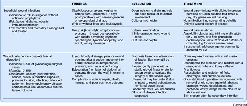
Wound and Abdominal Wall Infections
Superficial wound infections occur in up to 10% of patients who have undergone gynecologic surgery without perioperative antibiotics. The most common causes are Staphylococcus aureus and vaginal or enteric flora. The great majority of these infections are minor, although systemic toxicity or extensive infection may occur if the initial infection is neglected or in patients who are immunosuppressed, have diabetes, or are obese.
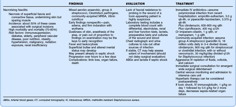
Thorough evaluation requires opening the wound for drainage and examination for deep fascial or muscular involvement. Superficial wound infections can be managed without antibiotics by meticulous wound care, irrigation with diluted hydrogen peroxide or Dakin solution four times per day, and dry gauze packing. Delayed wound closure can be performed if necessary. Table 125.2 details the clinical findings, evaluation, and treatment of wound infections, dehiscence, and necrotizing fasciitis.
Vaginal Evisceration
Vaginal evisceration (bowel and organs protruding from the vagina) is rare, yet dramatic and has a mortality of up to 10% because of associated intestinal necrosis, peritonitis, or other underlying or global illness. It occurs as a result of increased intraperitoneal pressure in the setting of a ruptured vaginal enterocele or unrecognized uterine perforation. The diagnosis is based on findings on physical examination.
Treatment
A moist covering must be placed immediately to protect the viscera. Bed rest in the supine or Trendelenburg position is recommended to prevent further outward pressure. Broad-spectrum antibiotics should be administered, and gynecology consultation should be obtained immediately for surgical repair.
Complications Specific to Laparoscopy
Laparoscopic procedures are characterized by more rapid recovery and lower complication rates than seen with open surgical procedures. However, unique complications are associated with needle or trocar insertion, induced pneumoperitoneum, and extensive use of electrocautery4,5 (Box 125.3). Most catastrophic complications are recognized intraoperatively. Management in the ED in the first month postoperatively is usually for wound complaints or symptoms caused by injury to the bowel, bladder, or ureters. Remote complications include hernias.
Box 125.3 Most Threatening and Most Common Complications of Laparoscopy Seen Postoperatively
Bleeding
Abdominal wall hematomas may occur as a result of damage to superficial vessels at laparoscopic sites. If the injury is extensive or if the size cannot be estimated because of habitus, a complete blood count can be obtained to estimate and track the blood loss, although the blood lost is not usually sufficient to require transfusion.

Full access? Get Clinical Tree

