1 Cardiovascular system
Anaesthesia and cardiac disease
ASSESSMENT OF RISK – Predictors in non-cardiac surgery
Cardiac complications are a major cause of perioperative morbidity and mortality, particularly vascular patients where mortality is 3–4% for open procedures. Perioperative MI accounts for 10–40% of all postoperative deaths. Risk stratification of patients with known, or at risk of, coronary artery disease is based on (1) the patient risk factors; (2) physiological status of the patient; (3) the risk factors of surgery.
Current thought is that if preoperative assessment reveals possible coronary artery disease, then patients booked for elective surgery should be referred to a cardiologist. Angioplasty in the lead up to elective surgery may increase overall 30-day mortality. CABG, however, may improve long-term outcomes in vascular surgical patients.
Previous ischaemia
Previous guidelines recommended waiting 6 months after an MI before non-cardiac surgery. It is now known that risk after an MI is related more to functional status of the ventricles and the amount of ischaemic myocardium. A small MI without residual angina, and good myocardial function, enables non-cardiac surgery 6 weeks post-MI. A large infarct, residual symptoms and LVEF <0.35 results in high risk even at 6 months post-MI.
Previous cardiac surgery/angioplasty
There is increased risk with non-cardiac surgery if the patient is <3 months post-CABG. Asymptomatic patients >6 months post-CABG are low risk.
Non-cardiac surgery performed within 6 weeks of PCI results in a high risk of stent thrombosis and infarction if the antiplatelet medication is stopped, or of major bleeding if the treatment is maintained throughout the operation.
Important risk studies
Hypertension
Diastolic blood pressure (DBP) is a good indicator of the severity of vascular disease. DBP >110 mmHg is associated with exaggerated swings in BP and an increased risk of perioperative complications. Severe (DBP >115 mmHg) or malignant (DBP >140 mmHg) hypertension should be treated before surgery. LV hypertrophy is associated with reduced ventricular compliance and these patients may benefit from perioperative monitoring of PCWP.
Perioperative hypertension doubles the risk of complications and is associated with increased silent ischaemia.
Investigations
Ambulatory ECG using 24 h Holter monitor. Ischaemic events are a highly significant predictor of adverse postoperative cardiac events. Silent preoperative ischaemia has a positive predictive value of 38% for postoperative cardiac events, whereas its absence precludes perioperative problems in non-vascular surgery in 99% of patients.
Exercise ECG (Bruce protocol). Aim for the target heart rate by stage 4. This is a good predictor of risk in patients with angina. Severe peripheral vascular disease limits exercising and may mask exercise-induced angina (consider the dobutamine stress test in these patients). ST-segment depression ≥0.1 mV during exercise is an independent predictor of perioperative ischaemic events.
ECHO. Ejection fraction, wall motion and valve abnormalities.
Thallium-201 scan. K+ analogue injected i.v. and taken up by well-perfused myocardium, showing underperfused areas as cold spots. Cold spots resolving by 4 h are areas of ischaemia; those persisting are infarcted tissue.
Technetium-99m scan. Similar to thallium scan but underperfused areas show as hot spots.
Dipyridamole–thallium scan. Dipyridamole causes coronary vasodilation to assess coronary stenosis. Similar effect with dobutamine, which also increases myocardial work, i.e. pharmacological stress test. Good predictor of postoperative cardiac complications.
Angiography. Definitive investigation. (Right coronary artery supplies sinoatrial node in 60% of patients and atrioventricular node in 50%). Indicated for unstable angina, or when there is a possible indication for coronary revascularization.
General anaesthesia for non-cardiac surgery
Choice of anaesthetic technique or volatile agent has no proven effect on cardiac outcome. Aim to optimize myocardial oxygen balance (Table 1.1).
Table 1.1 Factors affecting oxygen supply and demand
Supply | Demand |
---|---|
Coronary perfusion | Preload (LVEDP) |
O2 content | Afterload (SVR) |
Heart rate | Heart rate |
Contractility |
Premedication
Continue all cardiac medication until the day of surgery. Heavy premedication reduces anxiety, which may otherwise cause tachycardia, hypertension and myocardial ischaemia. Consider O2 after morphine premedication to avoid hypoxaemia from respiratory depression; the prevention of tachycardia results in less myocardial ischaemia overall.
β-blockade. Not all studies have shown benefit from perioperative β-blockade. The Peri-Operative Ischemic Evaluation (POISE) trial showed a beneficial effect of high-dose metoprolol on reducing the risk of perioperative MI, but at the risk of increased stroke and overall mortality. AHA 2007 guidelines recommend continuing β-blocker therapy in patients already on this medication, and giving β-blockers only to high risk vascular surgery patients.
Aspirin. Although aspirin increases the risk of bleeding complications, it does not increase the severity level of the bleeding complication. A meta-analysis has shown that aspirin withdrawal was associated with a three-fold higher risk of major cardiac events (Biondi-Zoccai et al 2006).
Monitoring
ECG. Leads II and V5 together detect 95% of myocardial ischaemic events. Leads II, V5 and V4R together detect 100% of events. ST segment monitoring may be a more sensitive indicator.
BP (invasive/non-invasive). Invasive BP monitoring enables blood gases/acid–base and K+ measurements.
CVP. Use the right atrium (RA) as zero reference point (midaxillary line, 4th costal cartilage). Normal range with spontaneous respiration is 0–6 cmH2O. The manubriosternal junction is 5–10 cm above the RA when the patient is supine. Ischaemia causes abnormal ‘v’ waves.
Pulmonary artery catheter. Good monitor of LV function but low sensitivity for detection of myocardial ischaemia (ischaemia causes ↑PCWP and ↑PAP). Rao et al (1983) showed increased reinfarction risk if preoperative PCWP was >25 mmHg. Thus, monitoring of PCWP and aggressive treatment with inotropes/vasodilators may reduce the risk of reinfarction. If ejection fraction >0.50 and there is no dyssynergy, CVP is an accurate correlate of PCWP, and PAP monitoring may be unnecessary.
Transoesophageal ECHO (TOE). Developed in the 1950s by Edler and Hertz. Ultrasound waves are formed when a voltage is applied across a substance with piezoelectric properties (usually lead-zirconate-titanate-5, PZT-5). Ultrasound waves are reflected back to the PZT-5 transducer, and converted back into electrical energy. This signal is then processed and displayed on a monitor. TOE requires less penetration than transthoracic ECHO and therefore uses a higher frequency (3.5–7 MHz) to produces higher resolution images.
Useful to assess perioperative:
Myocardial wall motion abnormalities detected by TOE are a much more sensitive method than ECG in detecting myocardial ischaemia. Post-bypass TOE is a sensitive predictor of outcome (MI, LVF, cardiac death).
Induction
Aimed at limiting hypotensive response to induction agent and hypertensive pressor response to intubation. High-dose opioid is a popular technique.
Anaesthetic
Avoid CVS changes that precipitate ischaemia. Tachycardia and hypertension increase myocardial O2 consumption and reduce diastolic coronary filling time. Hypotension reduces coronary perfusion pressure.
N2O is a sympathetic stimulant, but will decrease sympathetic outflow if the SNS is already stimulated, e.g. LVF. In the presence of an opioid, it may cause CVS instability.
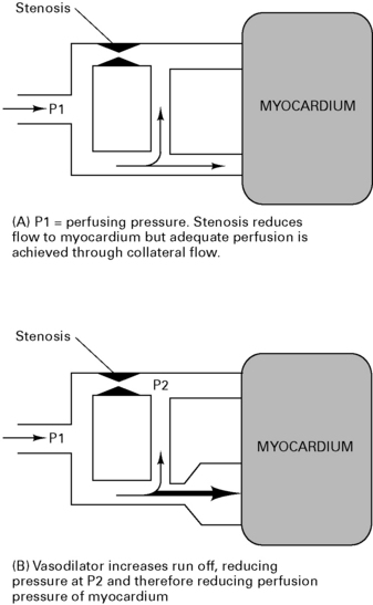
Volatiles. Enflurane and halothane both decrease coronary blood flow, but isoflurane, sevoflurane and desflurane increase coronary blood flow and maintain LV function in normotensive patients. Tachycardia with isoflurane increases myocardial work, but this is minimal with balanced anaesthesia. There is some concern that isoflurane may cause coronary steal (Fig 1.1) but it is thought not to do so as long as coronary perfusion pressure is maintained. There is growing evidence that isoflurane has myocardial protective properties, limiting infarct size and improving functional recovery. This mechanism mimics ischaemic pre-conditioning and involves the opening of ATP-dependent K+ channels causing vasodilation and preservation of cellular ATP supplies. Desflurane and sevoflurane probably have similar but less marked cardioprotective effects.
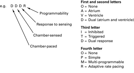
Figure 1.1 (A) Myocardial perfusion pressure (P1) reduced by stenosis. Adequate perfusion is achieved through collateral flow. (B) Vasodilator increases run-off, reducing pressure at (P2) and therefore reducing myocardial perfusion pressure distal to the stenosis.
Relaxants. Vecuronium combined with high-dose opioids tends towards bradycardia. Use of pancuronium avoids bradycardia.
Epidural/spinal
This decreases afterload and may improve LV function. General anaesthetic combined with epidural may cause severe hypotension because of vasodilation of vessels that have constricted above the block. In animals, redistribution of blood from epicardial to endocardial vessels reduces MI size. Angina following spinal anaesthesia tends to occur at cessation of the block, probably due to increased pre- and afterload aggravated by volume loading.
Blocks below L3 have no effect on the SNS. Blocks above T10 block sympathetic afferents to the adrenals and reduce catecholamine release. Blocks to T1–T4 interrupt cardioaccelerator fibres, preventing the coronary vasoconstrictive response to surgery, cause coronary vasodilation, decrease coronary perfusion pressure and decrease contractility and heart rate. Central hypovolaemia due to vasodilation causes a vagally mediated bradycardia which responds to fluid challenge.
Pacemakers
There are 200 000 patients with implanted pacemakers in the UK.
Intraoperative risks
Rate responsive pacemakers sense electrical activity or vibration around the pacing box and cause a tachycardia in response. Thus, shivering may cause a tachycardia. Fasciculations from suxamethonium are too transient to cause a tachycardia, but there is a case report of a pacemaker that stopped firing following administration of suxamethonium.
Pacemakers that sense blood temperature to control rate may trigger a tachycardia as a hypothermic patient is rewarmed. Those that measure respiratory rate by sensing thoracic impedance and adjust heart rate accordingly can also trigger a tachycardia if the ventilator is set at a high respiratory rate.
Diathermy
Diathermy current risks reprogramming the pacemaker (not AOO, VOO), causing microshock and inducing VF. Bipolar diathermy is the safest. If unipolar, mount the diathermy plate away from the pacemaker and use short bursts of minimum current. Do not use within 15 cm of the pacing box.
Application of a magnet over a non-programmable ventricular-inhibited pacemaker (VVI) reverts it to asynchronous mode (VOO). Application of a magnet over a programmable pacemaker increases the risk of reprogramming, but it will remain in an asynchronous mode until the magnet is removed, when the reprogrammed mode will take over. Do not use any magnets unless the pacemaker reprogrammes during surgery.
Automatic implantable cardioverter defibrillators (AICDs)
There are 4000 patients with implanted pacemakers in the UK, usually for drug-resistant malignant ventricular arrhythmias. This has reduced 1-year mortality from 66% to 9%. AICDs consist of a lead electrode system for sensing, pacing and delivery of shocks for cardioversion/defibrillation and a control unit consisting of a pulse generator, microprocessor and battery. Modern devices also act as DDD pacemakers.
Where the precise time since the onset of acute AF is uncertain, use oral anticoagulation for acute AF, as for persistent AF.
Atrial Fibrillation: the Management of Atrial Fibrillation
National Institute for Health and Clinical Excellence, June 2006
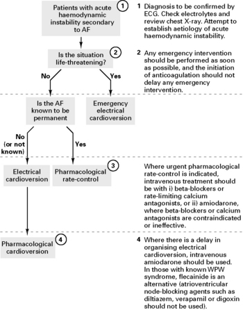
Antithrombotic therapy for acute-onset atrial fibrillation (AF)
For patients with acute AF who are receiving no, or only sub-therapeutic anticoagulation therapy:
For patients with a confirmed diagnosis of acute AF (<48 h since onset), use oral anticoagulation if:
Where a patient with acute AF is haemodynamically unstable, begin emergency treatment as soon as possible. Do not delay emergency intervention in order to begin anticoagulation treatment first.
Anaesthetic considerations for heart surgery
Aortic stenosis
Aortic stenosis becomes symptomatic when the normal valve area of 3 cm2 is reduced by >25%. Gradient >70 mmHg is severe (= 0.6 cm2); low cardiac output, dependent upon rate. Bradycardia reduces cardiac output and therefore BP. Tachycardia is poorly tolerated because reduced time for LV filling reduces ejection time and diastolic time during which coronary perfusion occurs. Aortic diastolic pressure must be maintained to preserve coronary blood flow. Ischaemia occurs even with normal coronaries. Atrial contraction is important to fill a poorly compliant LV. A fall in SVR is poorly tolerated. Therefore, maintain both pre- and afterload and avoid regional techniques.
Aortic regurgitation
This causes a dilated, overloaded and failing LV. Low aortic diastolic pressure impairs coronary perfusion. Slight tachycardia reduces regurgitant time and keeps LV small (Laplace’s law), thereby improving LV efficiency. A slight reduction in SVR reduces regurgitation but may reduce coronary perfusion pressure.
Mitral stenosis
A value area <1 cm2 is severe. There is poor LV filling and a fixed output, dependent upon rate. Low CO is worsened by tachydysrhythmia. Rapid heart rate reduces diastolic time for ventricular filling and thus reduces cardiac output, so bradycardia is beneficial. A high PVP risks pulmonary oedema with overtransfusion, but it is important to maintain adequate filling. A fall in SVR is poorly tolerated. Consider inotropes and pulmonary vasodilators.
Mitral regurgitation
Often well tolerated; PVP remains low. There is slight tachycardia and reduction in SVR which reduce regurgitation. Ischaemia is not usually a problem. General anaesthesia is normally well tolerated, unless pulmonary hypertension has developed, when inotropic support may also be indicated.
Hypertrophic obstructive cardiomyopathy (HOCM)
HOCM is autosomal dominant with variable penetrance. There is variable subaortic obstruction and impaired diastolic function. Try to avoid drugs that depress LV function. Increase pre- and afterload, maintain sinus rhythm and avoid excessive tachy/bradycardia. There is usually good LV function. Ventricular arrhythmias are common. Depression of myocardial contractility reduces outflow obstruction. β-agonists increase outflow obstruction.
Ischaemic heart disease
Rate pressure product (RPP) = heart rate × systolic pressure. Aim to maintain value at below 12 000.
Decrease preload (wall tension) and maintain afterload. A slight decrease in both contractility and rate can be beneficial by reducing work if there is good LV function. Myocardial depression may improve oxygenation unless there is severe IHD or aortic stenosis. If ischaemia occurs, decrease heart rate (β-blockers, calcium-channel blockers), increase ventricular volume (GTN) and increase afterload.
Pulmonary hypertension
Defined as mean pulmonary artery pressure (MPAP) >25 mmHg at rest. Overall, those with severe disease have a 5-year survival of only 27% from time of diagnosis, increasing to 54% with treatment.
Classification of pulmonary artery hypertension
Causes increased pulmonary vascular resistance (PVR) with increased load on the right heart causing right heart failure. Treatment aims to reduce PVR. Correct reversible causes (e.g. pulmonary thromboembolectomy, congenital heart disease, etc.). Pulmonary vasodilators include epoprostenol (prostacyclin) with its analogues (iloprost and treprostinil), endothelin receptor antagonists (bosentan), phosphodiesterase inhibitors (sildenafil) and inhaled nitric oxide.
Endocarditis prophylaxis
Antimicrobial Prophylaxis Against Infective Endocarditis in Adults and Children Undergoing Interventional Procedures
National Institute for Health and Clinical Excellence, March 2008 (http://www.nice.org.uk/nicemedia/pdf/CG64PIEQRG.pdf)
Summary
Antibacterial prophylaxis is not recommended for the prevention of infective endocarditis (IE) in patients undergoing procedures of the:
If patients at risk of endocarditis are undergoing a gastrointestinal or genito-urinary tract procedure at a site where infection is suspected, they should receive appropriate antibacterial therapy that includes cover against organisms that cause endocarditis.
Risk factors for the development of infective endocarditis

Full access? Get Clinical Tree

