Key Clinical Questions
When should a positive urine culture for Candida species be treated?
When should invasive candidiasis be suspected? How is it diagnosed and treated?
What is the clinical significance of a positive sputum culture for Aspergillus species?
How useful are antigen detection tests for the diagnosis of infection with Candida and Aspergillus species?
Introduction
Mucocutaneous infection with Candida species is common in the hospital setting, due to the high prevalence of diabetes and the use of broad-spectrum antibiotics. Invasive infections with Candida species were once rare, but have become much more common in hospitalized patients, particularly among those with compromised immunity, antibiotic exposure, and central venous catheters. The increased prevalence of Aspergillus infections in the hospital is almost wholly due to neutropenia from chemotherapy and the use of high-dose glucocorticoids and other immunosuppressive agents in patients with organ and bone marrow transplants and rheumatological diseases.
Candida Infections
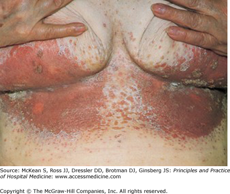
Candida species are normal flora of the oropharynx, bowel, and vagina. Candida overgrowth of these surfaces may arise in the setting of broad spectrum antibiotics, diabetes mellitus, or HIV infection, resulting in oral thrush, Candida esophagitis, and vaginal candidiasis. High estrogen states, such as pregnancy or oral contraceptive use, are an additional risk factor for vaginal yeast infection. Candida skin infections are often seen in moist, macerated intertrigenous folds, such as in the groin, perineum (diaper rash), pannus, axillae, and breasts.
While superficial candidiasis is common in both ambulatory and hospitalized patients, candidemia and disseminated candidiasis are usually seen in health care settings. Risk factors include critical illness, intensive antibiotic exposure, intraabdominal surgery, hemodialysis, central venous catheters, parenteral nutrition, intravenous drug use, and neutropenia.
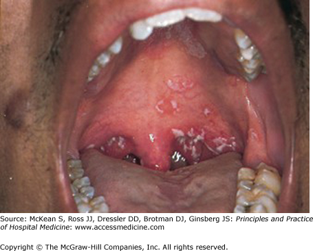
The diagnosis of mucocutaneous Candida infections is usually made clinically. Cutaneous candidiasis has a typical distribution in intertrigenous areas, with central erythema and maceration, surrounded by a collar of scale (Figure 192-1). Beyond this, there may be papular and pustular satellite lesions. Oral thrush (Figure 192-2) presents with painless white plaques on the mucous membranes and tongue that are easily scraped off. Fissures of the angles of the mouth (cheilitis) may also be present. Budding yeasts, hyphae, and pseudohyphae of Candida can be seen on microscopy when scrapings of skin or oral lesions are mixed with a drop of 10% potassium hydroxide, which digests host cells but not fungi.
Esophageal candidiasis presents with odynophagia. Oral thrush may, or may not, also be present. Endoscopy is required for confirmation in most patients. In AIDS patients with odynophagia, empiric treatment with fluconazole is reasonable. If symptoms do not subside in 3 to 4 days, endoscopy is indicated to exclude alternative causes such as herpes simplex, cytomegalovirus, and malignancy.
Vulvovaginal candidiasis presents with vaginal itching, soreness, dysuria, and dyspareunia. Vaginal discharge is often absent; when present, it may be thick and curdy, sometimes described by the patient as “cheesy.” Odor is usually absent. In addition to potassium hydroxide preparations, the usual investigations for the diagnosis of vaginitis should be performed, including the “whiff” test, vaginal pH measurement, and microscopy with saline (“wet prep”) for trichomonads, white cells, and clue cells. Sexually active women with vaginal discharge should also have studies to exclude chlamydial and gonorrheal infection.
Candida species are readily grown from urine culture. Most patients with candiduria have underlying illness, such as recent surgery, diabetes mellitus, an abnormal urinary tract, or malignancy, have been treated with antibiotics in the preceding month, and have indwelling urinary catheters or other urinary drainage devices. On many occasions when candiduria is documented, urine cultures were performed for inadequate reasons, such as cloudy urine without other symptoms. The vast majority of patients with candiduria are asymptomatic, and funguria tends to clear spontaneously without treatment. Progression to candidemia is unusual, and when it does occur, it is generally seen in patients who also have central venous catheters.
Candidemia and disseminated candidiasis (Candida infection of usually sterile sites) should be suspected in febrile or septic patients with exposure to antibiotics, intravenous catheters, parenteral nutrition, immunosuppressive agents, or chemotherapy. Candida species, unlike some other fungi, are not fastidious and can be grown without difficulty from blood cultures and subcultured onto regular blood agar plates. However, the diagnosis of disseminated candidiasis may be elusive as only half of patients with disseminated candidiasis have documented candidemia. The Candida score (Table 192-1) has been proposed as a diagnostic tool to define a population of sick patients at high risk of disseminated candidiasis who would benefit from early antifungal therapy.
| Risk Factor | Points |
|---|---|
| Multifocal candidal colonization | 1 |
| Recent surgery | 1 |
| Total parenteral nutrition | 1 |
| Severe sepsis | 3 |
The serum 1,3-β-D-glucan (Table 192-2) is a promising, if imperfect, adjunctive test in the diagnosis of invasive candidiasis. This component of the cell wall of Candida and other fungi may be detected in the serum of patients with candidemia and disseminated candidiasis, sometimes at high levels. However, the 1,3-β-D-glucan test currently lacks specificity, and false-positive results are common, especially for results close to the threshold of normal.
|
Patients with candidemia or possible disseminated candidiasis should be asked about focal complaints suggestive of metastatic Candida
Related posts:
 Strategies for Cost-Effective Care
 Building, Growing, and Managing a Hospitalist Practice
 Designing a Hospitalist Compensation and Bonus Plan
The Face of Health Care Emerging Issues for Hospitalists
Designing a Hospitalist Compensation and Bonus Plan
The Face of Health Care Emerging Issues for Hospitalists
 Medical Malpractice
Preventing and Managing Adverse Patient Events: Patient Safety and the Hospitalist
Medical Malpractice
Preventing and Managing Adverse Patient Events: Patient Safety and the Hospitalist

Full access? Get Clinical Tree


