 To assess for the absence of lung sliding, suggestive of a pneumothorax, in the following conditions:
 Blunt thoracoabdominal trauma
 Penetrating thoracoabdominal trauma
 Unexplained hypotension
 To assess for the presence of pleural fluid, suggestive of a hemothorax, in the following conditions:
 Blunt thoracoabdominal trauma
 Penetrating thoracoabdominal trauma
 Unexplained hypotension
CONTRAINDICATIONS
 If the EFAST examination delays a patient’s transport to the operating room
 Theoretical allergy to the ultrasound gel
ADVANTAGES
 Noninvasive
 No sedation required
 Performed at the bedside amidst simultaneous resuscitative efforts
 Does not require transportation to the radiology suite
 Serial examinations may be performed with changes in symptoms or hemodynamics
LANDMARKS
 Anterior Thorax
 The apical midclavicular line in the sagittal plane; transducer marker positioned cephalad
 Lateral Thorax
 The lateral thorax in the axillary region; transducer marker positioned obliquely and cephalad
 Pleural (Right: Hepatorenal and Left: Splenorenal)
 Transducer placed in the axillary line in the coronal plane at the level of 8th and 11th ribs; anterior axillary line on the right and posterior axillary line on the left, with the diaphragm as a landmark; transducer marker positioned toward the axilla.
TECHNIQUE
 The EFAST standard views in addition to the basic FAST examination:
 Bilateral anterior thorax
 Bilateral lateral thorax
 Bilateral pleural spaces
 Anterior Thorax
 Place the transducer in the second or third intercostal space in the midclavicular line
 The indicator should be cephalad
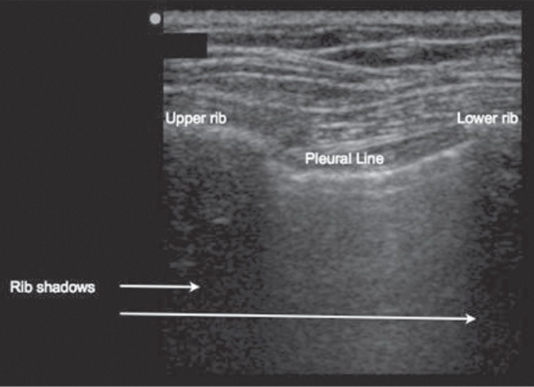
 Identify the bat sign: The upper rib–pleural line–lower rib profile (FIGURE 19.1)
 Normal lung findings
 B-mode: Visible sliding (shimmering or twinkling) at the level of the pleura
 B-mode: Comet tails—vertical reverberation artifacts arising from the pleural line (FIGURE 19.2)
 M-mode: Seashore sign (FIGURE 19.3)
 Pneumothorax
 B-mode: Loss of pleural sliding, as there is loss of contact between the visceral and the parietal pleura
 B-mode: Absence of comet tails
 M-mode: Stratosphere sign or bar-code sign (FIGURE 19.4)
 Lung point: Transition between collapsed and normally expanded lung; 100% specific for pneumothorax when identifiable
 TABLE 19.1 compares the signs suggestive of normal lung with those of pneumothorax



Full access? Get Clinical Tree


