 A subperichondrial hematoma that separates the perichondrium from the underlying auricular cartilage
 Result of a shearing force to the ear, but can also develop after blunt trauma
 The hematoma disrupts normal blood supply provided by the overlying perichondrium causing necrosis, fibrosis, and disfigurement of the auricle
 Commonly seen in wrestlers
 Early diagnosis and treatment is the key—necrosis begins within 24 hours
 In general, needle aspiration is sufficient. However, if a hematoma reaccumulates, incision and drainage (I&D) may be indicated.
CONTRAINDICATIONS
 There are no absolute contraindications
 Anticoagulation is a relative contraindication. Consult with ear, nose, and throat (ENT) specialist if there are any concerns.
RISKS/CONSENT ISSUES
 Risk of infection is low. If clinical suspicion is high, an antistaphylococcal antibiotic may be prescribed.
 Recurrent and untreated injuries allow new cartilage to develop, which leads to deformity of the auricle (cauliflower ear) (FIGURES 77.1 and 77.2)
LANDMARKS
 The pinna is most commonly involved
 The needle/scalpel is used in the area of greatest fluctuance
 General Basic Steps—Aspiration
 Cleanse
 Aspirate
 “Milk” hematoma
 Pressure dressing
TECHNIQUE
 For needle aspiration, a 10-mL syringe and a 20-gauge needle are required
 Cleanse the outer ear with a topical antiseptic (Betadine or chlorhexidine)
 Local anesthesia is seldom required. If used, avoid epinephrine (causes tissue necrosis).
 Stabilize pinna with thumb and fingers
 Puncture area of greatest fluctuance with needle
 “Milk” hematoma with thumb and index finger until completely evacuated
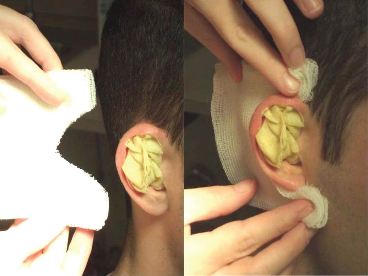
 Maintain pressure on ear for 3 minutes after needle has been withdrawn
 Apply antibiotic ointment and a pressure dressing
 Patient should be advised to avoid strenuous activity
 Check ear again in 24 hours for reaccumulation of hematoma
 Reaspiration may be required, whereas persistent reaccumulation warrants I&D



Full access? Get Clinical Tree


