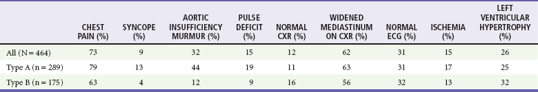
Chapter 85 Aortic dissection is a longitudinal cleavage of the aortic media created by a dissecting column of blood. The term “dissecting aortic aneurysm” has been inaccurately applied to this entity since 1819, when Laënnec first used the term aneurysme dissequant. The term aortic dissection is preferred to dissecting aortic aneurysm because the affected aorta is only rarely aneurysmal. In 1955, DeBakey outlined the principles that remain the basis for the surgical treatment of this entity. Medical treatment of aortic dissection was first advocated in the 1960s and is indicated for certain types of dissections.1–5 The in-hospital mortality rate for patients treated for aortic dissection is 27%.5 Aortic dissection occurs more often in men and increases with age.5,6 The incidence and prevalence are difficult to determine because of under-reporting of this condition. Mortality is 1 to 5 per 100,000 population per year. Hypertension is the most common risk factor associated with aortic dissection and is seen in most patients.5–7 A history of cardiac surgery is present in 18% and a bicuspid aortic valve in 14% of all patients with aortic dissections but more often in proximal dissections.5,7 Atherosclerosis is rarely involved at the site of dissection. Patients with aortic dissection may have a positive family history.8 Aortic dissection is uncommon before age 40 except in association with congenital heart disease, connective tissue disease, or inflammatory vasculitides. As many as 44% of patients with Marfan syndrome develop aortic dissection and account for about 5% of cases.5,7,9 Women with Marfan syndrome are at particular risk during pregnancy.10 In patients without connective tissue disease and with an aortic root size of less than 40 mm, pregnancy does not appear to be an independent risk factor.11,12 Loeys-Dietz syndrome is an autosomal dominant genetic syndrome associated with aortic aneurysms and skeletal features similar to Marfan syndrome. The vascular disease in these patients is aggressive, and the mean age of death is 26 years.13 Inflammatory vasculitides associated with thoracic aortic disease include Takayasu’s arteritis, giant cell arteritis, and Behçet’s disease.1 Acute aortic dissection also occurs with stimulant use, exertion, cardiac surgery, or intra-aortic balloon pump insertion.5 Blunt trauma from a high-speed deceleration injury usually causes traumatic aortic rupture, which is an entity distinctly different from aortic dissection (see Chapter 45). Medial degeneration, previously thought to be specific for aortic dissection, is now considered to be part of normal aging, although it is augmented by hypertension and with aortic dissection. The anatomic differences between the “normal” aorta and a dissection are quantitative rather than qualitative. Although medial degeneration was initially thought to be noninflammatory, more recent evidence suggests that inflammatory cell infiltration results from medial degeneration.14 The repetitive hydrodynamic forces produced by the ejection of blood into the aorta with each cardiac cycle contribute to weakening of the aortic intima and to medial degeneration. These hydrodynamic forces primarily affect the ascending aorta. Sustained hypertension intensifies these forces and results in an increase in medial degeneration. A bicuspid valve, present in 1 to 2% of the population, is the most common congenital abnormality affecting the aortic valve and proximal aorta.15,16 A bicuspid aortic valve may disrupt laminar flow and reorient the flow of blood toward the aortic wall, producing local injury. In Marfan and Ehlers-Danlos syndromes, normal hydrodynamic forces act on an aortic media that is already weakened. Anatomic classification is important for diagnosis and therapy. The Stanford classification is based on the involvement of the ascending aorta. Type A dissections involve the ascending aorta; type B dissections do not (Fig. 85-1). Dissections that involve the ascending aorta are more often lethal than those limited to the distal aorta and call for a different therapeutic approach. In the International Registry of Acute Aortic Dissection (IRAD), 62% of dissections are type A and 38% are type B.3–5 Patients with distal dissections tend to be older, heavy smokers with chronic lung disease and more often have generalized atherosclerosis and hypertension compared with patients who have proximal aortic dissections. Two other aortic conditions are closely related to aortic dissection: intramural hemorrhage (IMH) and penetrating aortic ulcer (PAU).17,18 Both groups of patients have clinical symptoms and management recommendations similar to those for patients with aortic dissection. An IMH is a contained hematoma within the aortic wall and occurs in about 10% of aortic dissections.5 Rupture of the vasa vasorum is believed to be the initial event. Penetrating atherosclerotic ulcers of the aorta occur in older hypertensive patients with evidence of coronary artery disease. Computed tomography (CT) shows a focal ulceration without dissection, most commonly in the distal descending aorta. The progression of penetrating ulcers results in progressive aortic enlargement with saccular and fusiform aneurysm formation. Patients can have both an intramural hematoma and a penetrating atherosclerotic ulcer.19 Pain is by far the most common presenting complaint, affecting more than 90% of patients.5,7,9 Most cases of painless aortic dissection are chronic in nature.7 The pain is usually excruciating, occurs abruptly, is most severe at onset, and is typically described as “sharp” more often than “tearing” or “ripping.”4 A family history of thoracic aortic disease may be reported.8 The location of the pain may help localize the dissection. Anterior chest pain is associated with the ascending aorta, neck and jaw pain with the aortic arch, pain in the interscapular area with the descending thoracic aorta, and pain in the lumbar area or abdomen with involvement below the diaphragm. Migration of the pain consistent with propagation of the dissection suggests aortic dissection but occurs in only 17% of cases.5 The onset of aortic dissection is often accompanied by visceral pain symptoms, such as diaphoresis, nausea, vomiting, lightheadedness, and severe apprehension. Syncope occurs early in aortic dissection in approximately 9% of cases and may be the sole presentation in some patients.5,9 It most often heralds dissection into the pericardium, causing pericardial tamponade, but may occur from transient interruption of blood flow to the cerebral vasculature. Other causes of syncope from aortic dissection are hypovolemia, excessive vagal tone, and cardiac conduction abnormalities. Patients with aortic dissection and syncope have a higher mortality.20 Neurologic symptoms such as focal weakness or change in mental status occur in up to 17% of cases.5,7,9 Aortic regurgitation occurs in up to 32% of patients and is more common with type A dissections.5,7 The murmur of aortic insufficiency may have a musical, vibrating quality with variable intensity, and congestive heart failure may develop. The patient with presumed aortic dissection should be examined carefully for findings that suggest hemorrhage into the pericardium or tamponade, such as jugular venous distention, muffled heart sounds, tachycardia, and hypotension. When the integrity of one of the branches of the aorta is compromised, the expected ischemic findings occur. Pulse deficits and discrepancies in blood pressure between limbs can be helpful if present but have a sensitivity of only around 30%.5,7,9,21 Usually these are present in the upper extremities and result from involvement of one or both of the subclavian arteries. Obstruction of one or both common iliac or superficial femoral arteries may produce pulse deficits in the lower extremities. Arterial obstruction may occur by either of two mechanisms. An intimal flap produced by the dissection may cover the true lumen of a branch vessel, or the dissecting hematoma may compress an adjacent true lumen. Frequent reexamination may detect transient pulse deficits. Neurologic findings are related to the site of blood flow interruption. Proximal dissections are a more frequent cause of strokes or coma. Stroke treatment with a fibrinolytic agent in the patient with aortic dissection can be fatal. Distal dissections occluding the anterior spinal artery commonly cause ischemic paraparesis or ischemic peripheral neuropathy.7 In up to 3% of cases, a proximal dissection can dissect into the ostium of a coronary artery, most frequently the right coronary artery, and cause an acute myocardial infarction (MI), usually inferior to posterior.5 Failure to identify the inciting aortic dissection with incorrect administration of a fibrinolytic agent occurs in about 0.1 to 0.2% of MIs.22 Distal extension of aortic dissections into the abdomen can cause mesenteric ischemia, renal failure, femoral pulse deficits, and lower extremity ischemia.23 Routine laboratory tests are of little value in the diagnosis of aortic dissection. The hemoglobin usually is normal or unchanged from the patient’s baseline. The leukocyte count is commonly mildly elevated. Recently, there has been increasing interest in the biochemical diagnosis of acute aortic dissection, including C-reactive protein, myosin heavy chain, soluble elastin, and D-dimer levels.24 D-dimer is the most readily available plasma marker and has the most diagnostic promise. Several authors have suggested that a negative D-dimer testing result makes a diagnosis of aortic dissection unlikely, but there is insufficient evidence to support use of a D-dimer as the sole screening test for aortic dissection, and 2010 guidelines from several major specialty societies recommend against this practice.1,25–31 Studies using a combination of a clinical pretest probability rule and D-dimer testing are needed.32 The electrocardiogram (ECG) is often useful in excluding MI; however, 15% of patients with aortic dissection may have ECG abnormalities suggesting ischemia.5 Proximal dissections that involve the right coronary artery may show an inferior wall MI, and the constellation of symptoms and signs (pain, diaphoresis, hypotension) may be difficult to distinguish from those associated with primary acute MI. The ECG typically shows left ventricular hypertrophy in 26%, reflecting long-standing hypertension. Other findings include nonspecific ST-T wave changes and prior Q wave infarction. No abnormalities are noted on the ECG in 31% of cases (Table 85-1).5 Table 85-1 Characteristics of Aortic Dissection from the International Registry of Acute Aortic Dissection4
Aortic Dissection
Perspective
Epidemiology
Principles of Disease
Pathophysiology
Classification
Clinical Features
Physical Examination
Diagnostic Strategies
Electrocardiography

Full access? Get Clinical Tree


Aortic Dissection
Only gold members can continue reading. Log In or Register to continue


