 Anorectal foreign body (FB) in a stable, cooperative patient, which is:
 Palpable by rectal approach
 Absence of a sharp edge
CONTRAINDICATIONS
 Obtain surgical consult immediately in following instances:
 Signs of perforation, obstruction, or severe abdominal pain
 Nonpalpable FB
 Broken glass in rectum
 Uncooperative or intolerant patient
 Lack of equipment necessary for retrieval
 General Basic Steps
 Determine type of FB
 Radiography
 Obtain necessary equipment
 Patient preparation
 Analgesia
 FB removal
 Assess for structural damage
KEY ELEMENTS OF HISTORY
 Ingestion (e.g., bones, toothpicks) versus rectal insertion
 Size and composition of FB
 Time of ingestion/insertion
 Attempts made to remove FB
 Assess for red flags—fever, abdominal pain, hematochezia
 Assess for sexual/physical assault, sexually transmitted disease (STD) risk
LANDMARKS
 Determine the orientation, location, and composition of the anorectal FB and, thereby the appropriate approach to removal by the following:
 Detailed history
 Consider radiography
 Kidney, ureter, and bladder (KUB) x-ray
 Chest x-ray for free air detection (if concerned about perforation)
 Physical examination, including digital rectal examination (DRE)
 Visualization of the anorectum is enhanced with the patient in the lateral decubitus position, lithotomy position, or prone with knees tucked into chest
TECHNIQUE
 Equipment
 Depends on the composition and locale of the FB but may include:
 Anesthesia/analgesia
 Light source
 Speculum (i.e., vaginal speculum or anoscope) or Parks retractor to improve visualization
 Ring and/or tenaculum forceps
 Foley catheter and/or endotracheal tube (ETT)
 Vacuum extractor
 Patient Preparation
 Get informed consent detailing risks, benefits, and alternatives
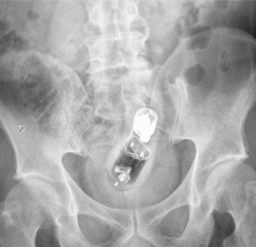
 Order KUB x-ray to localize and define FB, and to assess for obstruction or perforation if clinically necessary (FIGURE 83.1)
 Parenteral sedation and analgesia to enable relaxation and tolerance of the procedure. Avoid oversedation because the patient must be alert to assist in the delivery of the FB.
 Place the patient in the desired position
 A perianal block may facilitate further sphincter relaxation. This is achieved by superficial injection of local anesthetic (≤1.5 mg/kg of 0.5% bupivacaine or ≤7 mg/kg of 1% lidocaine with 1:100,000 epinephrine) in a ring around the anus.
 Examination
 External examination: Assess for signs of trauma
 DRE
DRE
 Gauge location and orientation of FB
 Assess for discharge or bleeding
 Small, blunt FBs may be removed during DRE
 Anoscopy
 Assess for mucosal injury
 Visualize FB
 Removal of FB
 Attempt delivery of the FB by applying suprapubic pressure in synchrony with the patient bearing down



Full access? Get Clinical Tree


