 Dislocated Ankle Joint
 Demonstrated on plain radiographs
 Clinically dislocated with neurovascular compromise
CONTRAINDICATIONS
 Open dislocations without neurovascular compromise may be better managed in the operating room for cleaning before reduction
 After one or two unsuccessful attempts at reduction, orthopedic consultation should be considered
RISK/CONSENT ISSUES
 Neurovascular damage may result from reduction attempt
 Closed reduction may be unsuccessful and operative repair may be required
 Risks of intravenous (IV) analgesia/sedation
 Risks of regional anesthesia
 General Basic Steps
 Patient preparation
 Obtain radiographs
 Analgesia/Sedation
 Reduce joint
 Check neurovascular status
 Immobilize joint
 Postprocedure radiographs
LANDMARKS
 The ankle joint is a modified saddle joint that comprises the distal fibula, tibia, and the talus bone of the foot
 Is a stable joint with strong ligamentous support
 Dislocations are a result of significant forces applied to the ankle and are often associated with fractures; isolated dislocations are uncommon
TECHNIQUE
 Preprocedure Examination
 Search for other injuries, especially if high-energy mechanism
 Check neurovascular status of the foot
 Get prereduction radiographs of dislocation (anteroposterior [AP], lateral, mortise views)
 If there is neurovascular compromise or tenting of the skin, perform immediate reduction before obtaining radiograph
 Try to ascertain the mechanism of injury
 Analgesia and Sedation
 Procedural sedation
 Regional analgesia
 Bier block
 Hematoma block
 Procedure
 Technique depends on type of dislocation but, in general, involves downward traction on heel while a force opposite to the direction of the dislocation is applied
 Flexion of the hip and knee to 90 degrees may aid reduction by relaxing the gastrocnemius–soleus complex
 If no assistant is available this can be accomplished by hanging the patient’s knee over the end of the bed
LATERAL DISLOCATION (FIGURE 67.1)
 Most common ankle dislocation seen in the emergency department (ED)
 Usually result of forced inversion of the foot
 Associated with malleolar or distal fibula fractures
 May be associated with rupture of the deltoid ligament
 Presents with foot laterally displaced with the skin very taut over the medial aspect of the ankle joint
 Technique
 Place one hand on the heel and the other on the dorsum of the foot
 Apply longitudinal traction to the foot
 While assistant applies countertraction to the leg, gently manipulate the foot medially. Successful reduction usually produces a palpable thud.
POSTERIOR DISLOCATION (FIGURE 67.1)
 Usually result of forced plantar flexion or a strong forward force applied to the posterior tibia
 Most are associated with a fracture of one or more malleoli
 Presents with the ankle held in plantar flexion with foot shortened in appearance and resistant to dorsiflexion
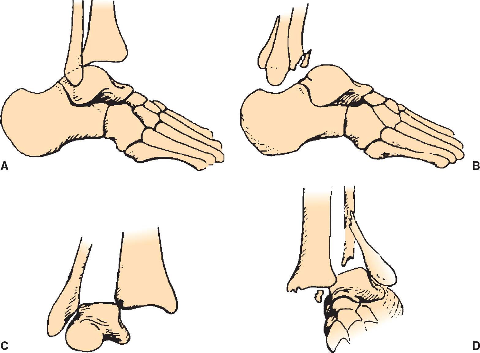
FIGURE 67.1 Four types of ankle dislocations. A: Posterior. B: Anterior. C: Superior. D: Lateral. (From Simon RR, Brenner BE. Emergency Procedures and Techniques. 4th ed. Philadelphia, PA: Lippincott Williams & Wilkins; 2002:285, with permission.)



Full access? Get Clinical Tree


