Partial thickness burns greater than 10 % TBSA.
Burns involving the face, hands, feet, genitalia, perineum, or major joints.
Third degree burns in any age group.
Electrical burns including lightning injuries.
Chemical burns.
Inhalational injuries.
Burn injuries in patients with preexisting medical disorders which could complicate management.
Combined burns and trauma.
Burned children in facilities that are not equipped to handle the care of children.
Burn injuries in those who will require social, emotional, or rehabilitative interventions.
Thermal injuries represent a special problem in terms of transporting patients to major burn centers. Thermal injuries, however, tend to occur away from centralized burn centers, most often in the rural setting. The first point of care for these patients then becomes smaller hospitals and clinics, in which practitioners may have limited experience in treating burned patients, and the estimation of the severity of burns is one of the key components of burn care, and determines the proper triage of patients. The combination of the relative rarity of encountering these injuries for the rural practitioner along with the development of centralized expertise results in a tendency towards both over- and under-triage of burned patients. At least one study demonstrated that 48 % of patients treated at a non-burn center meet criteria for transfer but were never transferred. The same study demonstrated that 12 % of patients treated at burn centers did not meet criteria for transfer [2]. Other studies have shown upwards of 30 % of transfers to burn centers not meeting criteria [3]. In an effort to optimize triage of burn patients to centralized centers from rural areas, several groups have looked to telemedicine as an initial tool in evaluation, prior to transfer.
Telemedicine and Triage of the Burned Patient
Telemedicine has been proposed as a solution to providing limited availability of clinical expertise in areas where it is not feasible to routinely have those clinicians on-site. The use is currently being explored in rural and international medicine, along with the possibility of aiding in disaster management [4–7]. Telemedicine technology is already being used in many burn centers in the USA. A survey submitted to directors of 126 burn center directors showed that 84 % of the 50 respondents were already using telemedicine in some form, with the majority using it routinely. While the majority of current telemedicine involves the use of remote patient data via home computer or personal devices, several reported the remote evaluation of acute burns for consultation, help in determining the need for transfer, and for follow-up visits [8].
While telemedicine has been thought of as an expensive solution to the problem of a lack of specialized clinical expertise in rural areas, the modern situation is such that most healthcare practitioners own or have access to common technology such as cellular phones and desktop computers which have free and user-friendly technology to be able to consult experts easily and quickly. The challenge for the current generation of practitioners will be ensuring the security of these interactions when utilizing this technology and compliance with the Health Insurance Portability and Accountability Act [9].
Initial Care of the Burned Patient
Acute care of the severely burned patient follows ATLS guidelines, ensuring that the patient has suffered no other significant traumatic injury, determination of the size of the patient’s burn, with potential transfer to a designated burn center. Burn injuries that are large enough to cause pathophysiologic responses such as hypotension and tachycardia warrant admission to the Intensive Care Unit. This response is secondary to intravascular fluid deficit within the first 24–48 h, and can lead to hypovolemic shock coupled with significant edema secondary to capillary leak. Early fluid administration in this group of patients has been shown to improve survival and decrease morbidity. While several formulas have been developed to estimate the fluid requirements to abate the associate morbidity and mortality, most centers utilize urine output as a measure of end-organ perfusion during this resuscitation, with a target output for most adults of 0.5 cc/kg/h.
Burn Resuscitation: Development of Closed-Loop and Decision-Assist Technologies
The logistics of how burn resuscitation actually occurs at most major burn centers involves practitioners using one of several available formulas (e.g., Parkland, Brooke Army) to determine a starting point for fluid volumes to be given to a severely burned patient. From that point on, most decisions are made either by institutional protocols driven by historical data, or clinical gestalt of the practitioners at the bedside. This frequently results in either fluid rates being reduced too quickly, or in the phenomenon of “fluid creep” whereby too much fluid is given in patients whose resuscitations are difficult. Studies have demonstrated that a vast majority of patients, in fact, are given more fluid than formulas estimate. The end product of either issue is a worse outcome for the patient. The need for automated systems has arisen out of these concerns, and allows for the skills and knowledge of expert practitioners to be utilized when they themselves are not available.
Personnel trained in burn care must provide prompt initialization of fluid therapy coupled with continuous attentive care at admission to the ICU, as described above. Appropriate fluid titration during this resuscitation has been one of the cornerstones of effective burn care; however, it is labor- and experience-dependent. Some groups have utilized computerized decision-support systems as early as 2007 [10]. These systems capture urine output volumes from a digital urimeter throughout the resuscitation, and have infused fluid volumes recorded and entered by bedside practitioners.
Computerized decision support system use has been shown to lower infused crystalloid volumes while maintaining urine output at goal a higher percentage of the time compared with traditional fluid resuscitation strategies. Furthermore, the use of this technology may also reduce ventilator free days and mortality in severely burned patients. While these tools have been developed to make the resuscitation of severely burned patients more standardized and hopefully safer, they are likely not a solution for a lack of expertise.
Determination of Burn Depth
Practitioners with expertise in burn wound management understand that the depth of a burn can be difficult to ascertain at its initial presentation. Determining which wounds would benefit most from excision and grafting versus a strategy of waiting for healing within 3 weeks remains one of the most important components of surgical planning for burned patients, and yet remains one of the most subjective. One of the developing technologies in the field of burns relates to being able to determine the precise depth of injury such as to preserve areas of skin that are still viable and/or to prevent unnecessary full-thickness excisions. The gold standard for assessing depth is a biopsy with histologic analysis. While it has been well studied as a reliable method for depth assessment, it is not always readily available, depending on local resources. Furthermore, it is largely impacted by sampling error, and as most burn wounds are not completely homogenous could lead to the need for multiple biopsies, increasing cost and possibly worsening the cosmetic outcome if the burn is not full-thickness.
As histologic analysis proves to be difficult to use in the clinical setting, the most commonly used method of determining burn depth currently is clinical evaluation. It is the least expensive method available, and relies on a subjective evaluation of the features of the burn wound, such as its appearance, sensitivity to touch, and pain [11–13]. However, none of the features of clinical appraisal of a burn have been shown to be reliable, and the accuracy has been called into question [14]. Studies have demonstrated, however, that this assessment is accurate in only 60–75 % of cases and that the clinical observation between examiners is not always consistent. Most frequently, the error is in depth overestimation [15].
Measurement of tissue perfusion has become an area of interest for groups examining technology to measure burn wound depths. Experimental data has demonstrated that the preservation of microvascular blood flow is related to the severity of a burn wound [16–18]. Technologies such as thermal imaging, vital dye use, and laser Doppler imaging are being considered for commercial use in determining burn depth.
Thermal imaging, or thermography, is a promising technology in that it is fast and noninvasive. It is based on temperature measurements of the wound to determine depth with the assumption that deep wounds are cooler by virtue of less or nonexistent vascular perfusion. Thermography has been criticized for falsely reporting deeper wounds secondary to ambient heat losses to the environment, and has been shown to have poor accuracy as wounds age [11].
Several variations of vital dyes have been utilized in research to differentiate between superficial and deep burns. The majority of these proved to have little clinical utility; however, some of these dyes have made a resurgence in many areas of medicine. Fluorescein fluorescence involves IV injection of fluorescein dye, followed by illumination via ultraviolet light over the area of interest. This has been utilized in burn wounds, however could not differentiate between deep and superficial burns, and had difficulty penetrating eschar [19, 20]. Another dye, Indocyanine green (ICG), is also administered intravenously, and has a peak spectral absorption at 800 nm. ICG binds to plasma proteins, becoming trapped in the vascular system, and is cleared with a half-life of 180 s via biliary excretion. This technology has been adapted to a videography image allowing practitioners to view this indicator of tissue perfusion on a monitor. Studies have confirmed that this allows for determining dermal viability from a structural and functional standpoint [21]. This technique is in use in several other fields of medicine via the SPY Elite® (LifeCell Corp, Branchburg, NJ) device and other similar technologies. Currently, the technology is being used in colorectal surgery, breast oncology, abdominal wall reconstruction, and vascular surgery/wound care. The most useful aspect of the SPY Elite is that it can be used in real-time, in an operating room, and can allow for sequential measurements in burned areas that are in question. While indocyanine green technologies appear promising, they are currently limited by availability and expense.
Laser Doppler imaging is the technology that has received the most attention thus far, and has a long list of studies supporting its use in burn wound assessment and treatment [14, 22, 23]. It has been used since the 1970s to evaluate cutaneous circulation. Doppler signals are produced when waves are reflected moving objects, and measured by the change in frequency based on the space between the object and the transducer. Such techniques have been examined in burns and demonstrated the highest positive predictive values of any of the technologies available currently [24]. As such, it is the only technology which has been FDA approved for burn wound assessment. It has consistently been shown to be better than clinical assessment at multiple time points following injury [23].
Management of the Burn Wound
Burn Eschar Excision
Debridement of burn eschar typically involves sharp dissection using instruments such as Bovie electrocautery, scalpels, and Humby knives. These methods of debridement are swift and effective; however, healthy collateral tissue around the wound may be removed as well. Furthermore, while experienced burn surgeons can typically perform this easily for full-thickness burns, superficial and partial-thickness burns do not need such deep excision, and sharp dissection may result in a much deeper excision than is necessary. Blood loss can also be an issue when very large surface areas are involved. To reduce unnecessary excision depth and size, more sophisticated technology is being developed for excision of burn eschar, especially for the case of partial-thickness burns. One example, the Versajet™ (Smith & Nephew, London, UK) system, uses pressurized sterile saline which is forced into a specialized nozzle which focuses the liquid into a jet. This jet is passed along tissue and takes with it ablated tissue and debris, moving the mixture into an evacuator port. This system has been shown to reduce bacterial burden and reduces the volume of normal tissue excised. Further examination of this and similar excision technologies will delineate the patients that most benefit from this form of debridement.
The Skin Bank
One of the most important and advanced technologies stemming from the care of thermally injured patients is the development of the Skin Bank. While skin grafting techniques developed in the 1800s, it was not until the early 1900s that the storage of human skin began. Early on, skin could be stored for up to 2 weeks prior to use. By the 1940s, the use of allograft skin in burn wound coverage had been described in children with extensive burns [25]. Finally, in 1949, the United States Navy Tissue Bank was established, heralding the era of the modern Skin Bank. Soon after, the American Association of Tissue Banks and the Food and Drug Association set policies and standards for tissue harvesting, processing, and banking of human tissues.
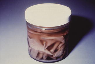
The technical aspects of skin banking include a rigorous donor screening process, followed by skin recovery within 24 h of the death of the donor. The skin is then processed in a laminar flow clean room. This processing involves microbiologic testing for bacteria, yeast, and fungi. These tests are performed on multiple samples of skin from each 10 % of the body surface area from which the skin was removed. Fresh allograft skin is then stored at 4 °C in tissue culture medium. This typically allows for storage for up to 7–10 days, after which the skin is cryopreserved (Fig. 16.1).
 End Points of Resuscitation
 Cardiac Surgery Advances: Do We Still Remember How to Do the Open Bypass?
 The Role of Robotics in Selective Thoracic Surgical Problems: Technical Considerations
 Artificial Limbs for Upper Extremity Amputation
 Use of Biologic Grafts in Surgery
 Brain Cancer: The New Frontiers


Fig. 16.1
Fresh allograft skin prior to freezing. Fresh skin can be used without freezing which many claim to have better results through better graft take
Related posts:
End Points of Resuscitation
Cardiac Surgery Advances: Do We Still Remember How to Do the Open Bypass?
The Role of Robotics in Selective Thoracic Surgical Problems: Technical Considerations
Artificial Limbs for Upper Extremity Amputation
Use of Biologic Grafts in Surgery
Brain Cancer: The New Frontiers
Full access? Get Clinical Tree
