Fig. 50.1
Neonatal extracorporeal membrane oxygenation (ECMO) circuit. The arrows show the direction of blood flow
There have been substantial technological improvements in contemporary ECMO support devices. Modern hollow-fiber oxygenators provide much lower resistance across the oxygenator membrane, allowing for the use of centrifugal pumps in the ECMO circuit. Centrifugal blood pumps are being used with increased frequency over traditional roller pumps. Both magnetically driven and magnetically suspended centrifugal pumps are available and are designed to reduce mechanically induced hemolysis. Some of the newest pumps can even provide pulsatile blood flow. Newer cannula designs, such as the double-lumen Avalon cannula (Avalon Elite®; Avalon Laboratories, Rancho Dominguez, CA), allow for more effective circuit flow and decrease recirculation during VV-ECMO support. Ongoing research efforts are directed toward improving the biocompatibility of circuit surface-blood interfaces in hopes of reducing the need for anticoagulation.
Minimally Invasive Surgery
Minimally invasive surgical (MIS) techniques have proven safe and efficacious in infants, children, and adolescents. Their main advantages relate to decreased postoperative pain, reduced hospital stay, faster return to normal activities, and superior cosmetic results when compared to conventional open techniques. Disadvantages include loss of haptic feedback, steeper learning curves, and increased operative costs for many procedures. Driven by numerous influences, MIS has quickly become the standard of care for many pediatric surgical conditions.
Laparoscopy
In 1987, Philippe Mouret reported the first laparoscopic cholecystectomy in Lyon, France. Since that historic event, the use of laparoscopy in general surgery has grown exponentially. This worldwide revolution has been driven by patient demand, surgeon ingenuity, and advances in medical technology. Initially, pediatric surgeons were slow to embrace laparoscopic techniques. Many believed that they were already performing most procedures through small incisions, and their patients recovered quickly. Optics and instruments were designed for adult patients, and their use in children was often cumbersome.
In the past two decades, technological advances have produced progressively smaller laparoscopic instruments and higher quality imaging. Smaller, lightweight trocars have been specifically designed for pediatric patients. Rigid laparoscopes (3 and 5 mm diameters) with angled lenses (30°, 45°, and 70°) foster the ability to work in confined spaces. Complete 3 and 5 mm instrument sets with shorter lengths have been engineered to suit the needs of pediatric surgeons. Advanced energy devices, such as the LigaSure™ vessel sealing system (Covidien, Mansfield, MA) and the Harmonic ACE® (Ethicon Endo-Surgery, Cincinnati, OH), achieve hemostasis and tissue dissection with minimal lateral thermal spread or tissue damage.
Today, advanced pediatric laparoscopic techniques have been applied to virtually every general and thoracic procedure in children. Laparoscopy is routinely used for cholecystectomy, appendectomy, pyloromyotomy, splenectomy, adrenalectomy, small bowel resections, exploration, and more. Advanced procedures include ileocolectomy for Crohn’s disease, endorectal pull-through for Hirschsprung’s disease, resection of choledochal cyst, and Roux-en-Y gastric bypass. Thoracoscopy has become standard for decortication, wedge resections, and minimally invasive repair of pectus excavatum. More advanced procedures include lobectomy, tracheoesophageal fistula repair, and congenital diaphragmatic hernia repair (Fig. 50.2).
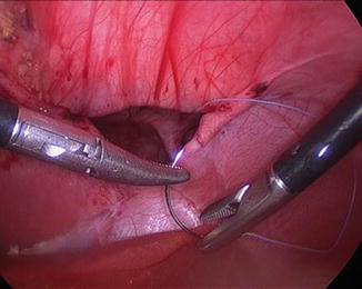
Fig. 50.2
Minimally invasive repair of a Morgagni diaphragmatic hernia in an infant
Recently, there has been increasing enthusiasm about decreasing the number of ports used for laparoscopic and thoracoscopic surgery. The most common techniques are single-port and single-incision laparoscopic surgery (SILS). The development of articulating cameras and instruments has made these techniques more plausible by allowing spatial separation and improved optics. Modified single ports are available which measure a few centimeters in diameter and allow the introduction of the scope and two instruments. In SILS, two or three small trocars are placed through an umbilical incision. There are successful reports of cholecystectomy, appendectomy, splenectomy, intussusception reduction, gastrostomy tube placement, thoracoscopic lung biopsy, decortications, and other pediatric procedures. The main advantage of these techniques is improved cosmetic results. Disadvantages include increased postoperative pain and incisional hernias.
Robotic Surgery
Robotic surgery is an emerging technology that may expand the variety of MIS procedures that can be accomplished in pediatric patients. Advantages of robotic surgery are its greater surgical precision, articulating instruments with increased range of motion, enhanced 3-D high-definition visualization of the operative field, reduced tremor, and improved ergonomics for the surgeon. Disadvantages include extended operative times, prolonged exposure to anesthesia, and significant expense of new devices, software upgrades, and operating room costs. Because of loss of domain, robotic surgery is difficult to perform in infants weighing less than 4 kg.
Essentially, any procedure can be done robotically. It is debated as to which procedures should be performed robotically and which procedures are more appropriate for other MIS techniques. Robotic techniques are best suited for complex operations which require fine dissection and suturing. Abdominal procedures include fundoplication, gastric bypass, resection of tumors, colectomy, and repair of congenital defects such as duodenal atresia, gastric duplications, and diaphragmatic hernia. Thoracic procedures include lobectomy, segmentectomy, tracheoesophageal fistula repair, and resection of mediastinal masses. Genitourinary and pelvic procedures include pyeloplasty, extramucosal ureteral reimplantation, and ovarian teratoma resection.
Fetal Surgery
Fetal surgery is a developing field. In the past three decades, it has evolved from a fanciful idea to a dedicated medical subspecialty. Fetal surgery is conducted in specialized centers throughout the USA and Europe. The fetal surgeon is an integral part of a collaborative, multidisciplinary fetal care team. Health care professionals from the fields of obstetrics, perinatology, genetics, neonatology, anesthesiology, radiology, and surgery must be readily available. Advances in fetal imaging and diagnosis have allowed clinicians to accurately classify congenital anomalies and stratify patients into appropriate risk groups. Each family must undergo extensive counseling that includes a detailed discussion of the maternal and fetal risks and potential benefits of a fetal intervention. Complications from fetal interventions include fetal demise, preterm labor, premature rupture of membranes, chorioamnionitis, and bleeding. Open fetal surgeries can cause uterine rupture in the current pregnancy and any future pregnancies. Procedures should only be offered when there is clear benefit to the fetus.
Congenital Lung Lesions
Congenital lung lesions (CLL) are a diverse group of malformations. Their antenatal detection has increased with the liberalized use of screening ultrasonography. These lesions are thought to exist across a spectrum of developmental pathways. Follow-up assessment with high-resolution ultrasound or ultrafast MRI is encouraged to better assess theses masses. The prenatal diagnosis and management of these lesions can be challenging, and urgent fetal intervention may be warranted in some cases.
Congenital cystic adenomatoid malformation (CCAM) is the most common CLL. A CCAM is a multicystic mass of nonfunctional pulmonary tissue that is usually confined to one lobe of the lung. The cysts can range in size from <1 mm to >10 cm. CCAMs can be classified as being macrocystic (>5 mm diameter for single or multiple cysts) or microcystic. However, prognosis depends on the size of the CCAM and its growth characteristics. CCAMs display unpredictable growth patterns between 18- and 26-week gestation. Thereafter, the growth rates plateau and may even regress.
Bronchopulmonary sequestration (BPS) is an area of nonfunctional lung tissue that does not communicate with the bronchial tree. It has a systemic arterial blood supply and variable venous drainage. An intralobar sequestration is located within the lung parenchyma. An extralobar sequestration is enveloped in its own pleura and may be located above or below the diaphragm.
Fetal hydrops is defined as the accumulation of fluid in two body cavities (pleural effusion, ascites, skin/scalp edema). Hydrops is often a harbinger of poor fetal or postnatal outcomes. The CCAM volume ratio (CVR) has emerged as a reliable way of predicting outcomes in a fetus with a CLL. CVR is calculated by measuring three tumor dimensions (length, width, height), multiplied by a constant (0.52), and divided by head circumference (to correct for gestational age). In a recent study, 56 % of fetuses with a CVR >2 required a fetal intervention, whereas only 3 % of fetuses with a CVR <2 required intervention [7]. Smaller CLLs and larger CLLs without hydrops can be followed with weekly or bimonthly ultrasound studies.
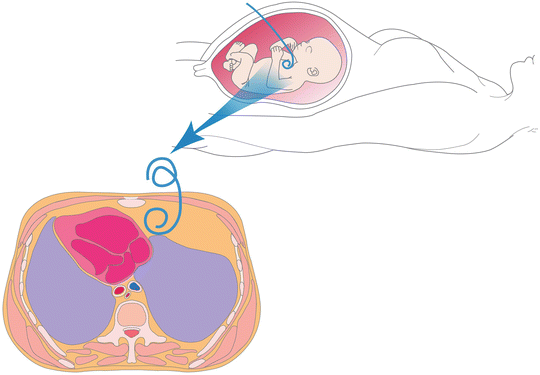
Fetal interventions are reserved for only the most severe CLLs. Prenatal steroids should be considered in patients with large, symptomatic solid or microcystic lung masses. Lesion regression and hydrops reversal have been reported from multiple centers. Ultrasound-guided thoraco-amniotic shunting can be attempted in fetuses with large macrocysts or BPS-related severe pulmonary effusions (Fig. 50.3). Open fetal surgery is reserved for cases where fetal demise is imminent. These procedures are considered to patients younger than 30-week gestation with predominantly solid lesions and hydrops. Fetal lobectomy can be performed, but preterm delivery remains a challenge. In patients greater than 30 weeks, an ex utero intrapartum therapy (EXIT) procedure may be considered. An airway is secured and the lung mass is resected while the patient remains on placental bypass.
 End Points of Resuscitation
 Cardiac Surgery Advances: Do We Still Remember How to Do the Open Bypass?
 The Role of Robotics in Selective Thoracic Surgical Problems: Technical Considerations
 Artificial Limbs for Upper Extremity Amputation
 Use of Biologic Grafts in Surgery
 Brain Cancer: The New Frontiers


Related posts:
End Points of Resuscitation
Cardiac Surgery Advances: Do We Still Remember How to Do the Open Bypass?
The Role of Robotics in Selective Thoracic Surgical Problems: Technical Considerations
Artificial Limbs for Upper Extremity Amputation
Use of Biologic Grafts in Surgery
Brain Cancer: The New Frontiers
Full access? Get Clinical Tree
