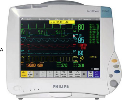
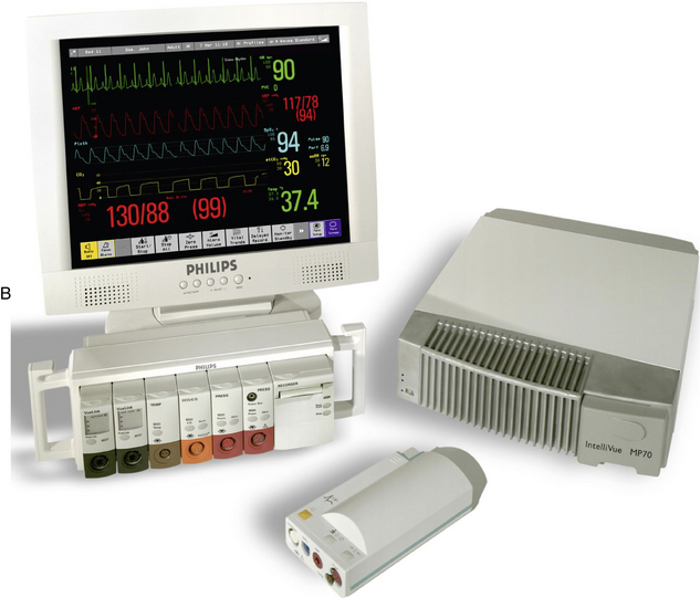
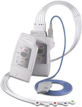
PROCEDURE 57 • Knowledge of the anatomy and physiology of the cardiovascular system, principles of cardiac conduction, principles of electrophysiology, electrocardiogram (ECG) lead placement, basic dysrhythmia interpretation, and electrical safety is necessary. • Advanced cardiac life support knowledge and skills are needed. • Electrophysiologic monitoring with hardwire and telemetry is indicated for all patients in critical care units and for patients in selected acute care settings, postanesthesia areas, operating rooms, and emergency departments. • Electrophysiologic monitoring is designed to give a graphic display of the electrical activity in the heart generated by depolarization and repolarization of cardiac tissue. • Hardwire ECG monitors have electrodes and lead wires that are attached directly to the patient. Impulses are transmitted directly from the patient to the monitor (Fig. 57-1). • Telemetry systems have electrodes and lead wires that are attached from the patient to a battery pack and transmit impulses to the monitor via radio wave transmission (Fig. 57-2). • Telemetry is useful in progressive ambulation and evaluation of activity tolerance. A disadvantage to telemetry is that ambulation and activity can increase distortion of the ECG pattern. • Specific areas of the chest are used for electrode placement to obtain a view of the electrical activity in a particular area of the heart (commonly called a lead). • ECG monitors use a three-lead or five-lead wire system to provide different views (leads) of the heart’s electrical activity. Most acute and critical care units use a five-lead system for continuous bedside monitoring or telemetry. • Standardized placement of leads is important so that information obtained is assessed within a common frame of reference and so that appropriate judgments can be made on the patient’s cardiac status. Alterations of electrode position may distort the appearance of the waveform significantly and can lead to misdiagnosis or mistreatment. • The two major factors that determine the views of the ECG deflection on the monitor are the location of the electrodes on the body and the direction of the cardiac impulse in relation to the position of the electrode. • A basic rule of electrocardiography is the rule of electrical flow. This rule notes that if electricity flows toward the positive electrode, an upright pattern is produced on the monitor or graph paper. If the electricity flows away from the positive electrode (or toward the negative electrode), a downward pattern or deflection is produced on the monitor or graph paper. Lead wires attached to the patient are coded (+, P [positive]; or –, N [negative]; RA [right arm]; RL [right leg]; LA [left arm]; LL [left leg]; V or C [V or precordial vector and C or chest lead] in some way for ease in correct placement. Placement of the leads gives different views of the electrical conduction through the heart. • Information from the bedside via hardwire or telemetry can be transferred to a central monitor, where it can be printed, stored, and analyzed (Fig. 57-3). • Five-lead bedside monitoring systems provide a continuous readout of two or more leads simultaneously. This readout provides more information and a comparison of the ECG patterns. Optimal lead selection is based on the goals of monitoring for each patient’s clinical situation.
Electrocardiographic Leads and Cardiac Monitoring
PREREQUISITE NURSING KNOWLEDGE
Related posts:
![]() 39: Automated External Defibrillation
39: Automated External Defibrillation
![]() 139: Calculating Doses and Flow Rates and Administering Continuous Intravenous Infusions
139: Calculating Doses and Flow Rates and Administering Continuous Intravenous Infusions
![]() 16: Continuous Venous Oxygen Saturation Monitoring
16: Continuous Venous Oxygen Saturation Monitoring
![]() 20: Chest Tube Placement (Perform)
20: Chest Tube Placement (Perform)
![]() 95: Lumbar Puncture (Perform)
95: Lumbar Puncture (Perform)
![]() 35: Invasive Mechanical Ventilation (Through an Artificial Airway): Volume and Pressure Modes
35: Invasive Mechanical Ventilation (Through an Artificial Airway): Volume and Pressure Modes

Full access? Get Clinical Tree


57: Electrocardiographic Leads and Cardiac Monitoring