Key Concepts
-
•
Many orthopedic injuries can be stabilized and treated definitively by the emergency clinician. Consultation with an orthopedist should be sought for the treatment of some long bone fractures, open fractures, injuries with joint violation, tendon injuries, and injuries with neurovascular compromise.
-
•
A careful history and physical examination can predict radiographic findings in orthopedic injuries with a high degree of accuracy.
-
•
Open fracture management should focus on the early administration of antibiotics, tetanus prophylaxis, dressing coverage of the wound, and splinting. Suggested therapy for open fractures includes a first-generation cephalosporin, with the addition of an aminoglycoside for type II or III fractures.
-
•
Compartment syndrome is associated most commonly with a closed long bone fracture of the tibia but also is well described in the thigh, forearm, arm, hand, and foot and can occur with isolated soft tissue trauma. Clinical examination remains the diagnostic cornerstone of acute compartment syndrome, which may then be confirmed by compartment pressure measurement.
-
•
Because of the anatomic location and blood supply distribution, certain bones may undergo avascular necrosis after fracture, especially if fractures are comminuted and go untreated for any length of time. The femoral head, talus, scaphoid, and capitate are particularly prone to this complication.
-
•
Fat embolism syndrome is a serious consequence of fractures, occurring most commonly after long bone leg fractures in young adults and after hip fractures in older patients. Acute respiratory distress syndrome is the most serious manifestation. Neurologic involvement, manifesting as restlessness, confusion, and deteriorating mental status, as well as thrombocytopenia resulting in a petechial rash are early signs.
-
•
Patients should be evaluated for the ability to ambulate safely prior to discharge. The use of a walker or other device, if it can be used safely, is acceptable. It also must be assured that the patient or a caregiver can carry out essential activities of daily living.
Overall Foundations
Background and Importance
Orthopedic emergencies and musculoskeletal complaints and conditions comprise approximately 20% of all emergency department (ED) visits. Although rarely life-threatening, orthopedic injuries may threaten a limb or its function, and accurate early diagnosis and treatment can avert long-term complications. Outcomes, including preventable complications, may affect a person’s quality of life. Many orthopedic injuries can and should be treated definitively by the emergency clinician. Consultation with an orthopedist should be sought for the treatment of most long bone fractures, open fractures, injuries with joint violation, tendon injuries, and injuries with neurovascular compromise.
When evaluating a potential orthopedic injury, the following basic general principles should be considered:
-
1.
Most orthopedic injuries can be predicted by considering the chief complaint, age of the patient, mechanism of injury, and estimate of the amount of energy transferred during the injury.
-
2.
A careful history and physical examination can predict radiographic findings with a high degree of accuracy. A presumptive diagnosis before a radiographic study may prompt the emergency clinician to order special views necessary to diagnose an injury correctly. Many fractures were accurately described even before the advent of radiology ( Table 41.1 ).
TABLE 41.1
Common Fracture Names and Their Origins
Fracture Eponym or Name Description Comment Aviator Vertical fracture of the neck of the talus with subtalar dislocation and backward displacement of the body First described in flyers during World War I; arises from forced dorsiflexion of the foot in flying accidents and in traffic accidents after a head-on collision Barton Intraarticular fracture-dislocation of the wrist Considered complicated and unstable; requires surgical reduction in most cases; described by Barton in 1838 before the advent of radiography Dorsal Barton Oblique intraarticular fracture of the dorsal rim of the distal radius with displacement of the carpus along with the fracture fragment Results from high-velocity impact across the articular surface of the radiocarpal joint, with the wrist in dorsiflexion at the moment of impact Volar Barton Wedge-shaped articular fragment sheared off the volar surface of the radius (volar rim fracture), displaced volarly along with the carpus Similar mechanism as dorsal Barton but with wrist in volar flexion at time of injury; also referred to as reverse Barton’s fracture; much rarer than dorsal Barton fracture Bennett Oblique fracture through base of the first metacarpal, with dislocation of the radial portion of the articular surface Usually produced by direct force applied to the end of the metacarpal; dorsal capsular structures disrupted by the dislocation; marked tenderness along medial base of thumb Bosworth Fracture-dislocation of the ankle resulting in the fibula being entrapped behind the tibia Rare injury, produced by severe external rotation force applied to the foot; physical examination reveals foot severely externally rotated in relation to the tibia Boxer Fracture of the neck of the ring or small finger metacarpal Results from striking a clenched fist into an unyielding object, usually during an altercation, or against a wall, out of frustration or anger Chance Vertebral fracture, usually lumbar, involving the posterior spinous process, pedicles, and vertebral body Caused by simultaneous flexion and distraction forces on the spinal column, usually associated with use of lap seat belts; anterior column fails in tension, along with the middle and posterior columns; may be misdiagnosed as a compression fracture Chauffeur Solitary fracture of radial styloid Occurs from tension forces sustained during ulnar deviation and supination of the wrist; name derived from occurrence in chauffeurs who suffered violent, direct blows to the radius incurred while turning the crank on a car, only to have it snap back, during previous eras Clay shoveler Fracture of the tip of the spinous process of the sixth or seventh cervical vertebra First described in Australian clay shovelers who sustained a fracture of the spinous process by traction as they lifted heavy loads of clay Colles Fracture of the distal radius with dorsal displacement and volar angulation, with or without an ulnar styloid fracture Most common wrist fracture in adults, especially in older adults; results from fall on an outstretched hand; also known as silver fork deformity, which accurately describes the gross appearance in the lateral view; first described by Colles in 1814, before the advent of radiography Cotton Trimalleolar fracture Fracture of the lateral malleolus, fracture of the posterior malleolus, and either a fracture of the medial malleolus or disruption of the deltoid ligament, with visible widening of the mortise on ankle radiograph Dashboard fracture Fracture of the posterior rim of the acetabulum Named for mechanism of injury—a seated passenger striking the knee on a dashboard, driving the head of the femur into the acetabulum Dupuytren Fracture-dislocation of the ankle Results from a similar mechanism as the Maisonneuve fracture (i.e., external rotation of the ankle), resulting in deltoid ligament rupture or medial malleolus fracture, diastasis of the inferior tibiofibular joint, and indirect fracture of the fibular shaft; Maisonneuve was a student of Dupuytren Essex-Lopresti Fracture of radial head with dislocation of the distal radioulnar joint Results from longitudinal (axial) compression of the forearm Galeazzi Fracture of the shaft of the radius with dislocation of the distal radioulnar joint; ligaments of inferior radioulnar joint ruptured, head of ulna displaced from ulnar notch of the radius Results from fall on outstretched hand, with the wrist in extension and the forearm forcibly pronated; inherently unstable, with tendency to redisplace after reduction Hangman Fracture-dislocation of atlas and axis, specifically of pars interarticularis of C2 and disruption of C2–3 junction; separation occurs between second and third vertebral bodies from anterior to posterior side Results from extreme hyperextension during abrupt deceleration; most common cause is the forehead striking the windshield of a car during a collision; a bit of a misnomer in that hanging usually produces death by strangulation rather than cord damage Hume Fracture of the proximal ulna associated with forward dislocation of the head of the radius Essentially high Monteggia injury Jefferson Burst fracture of ring of C1, or atlas Axial loading results in a shattering of the ring of the atlas; decompressive type of injury; associated with disruption of transverse ligament; unstable injury Jones Transverse fracture of the fifth metatarsal base, occurring at least 15 mm distal to the proximal end of the bone, distal to the insertion of the peroneus brevis Should not be confused with the more common avulsion fracture of fifth metatarsal styloid, produced by avulsion at the insertion of the peroneus brevis; Jones described the fracture that bears his name in 1902, after sustaining the injury himself while dancing. Le Fort Maxillary fracture Types I, II, and III (see Chapter 34 ) Le Fort-Wagstaffe Avulsion fracture of the anterior cortex of the lateral malleolus Rare pull-off injury of the fibular attachment of the anterior tibiofibular ligament Lisfranc Fracture located around the tarsometatarsal (Lisfranc) joint, usually associated with dislocation of this joint Lisfranc, a field surgeon in Napoleon’s army, described an amputation performed through the tarsometatarsal joint in a soldier who caught his foot in a stirrup when he fell off his horse; since then, the joint has borne his name. Maisonneuve Fracture of proximal third of fibula associated with rupture of the deltoid ligament or fracture of the medial malleolus and disruption of the syndesmosis Results from external rotation of the ankle with transmission of forces through syndesmosis; proximally, the force is relieved by fracture of the fibula; described experimentally in 1840, before radiography Malgaigne Fracture of the ilium near the sacroiliac joint with displacement of the symphysis, or a dislocation of the sacroiliac joint with fracture of both ipsilateral pubic rami Resultant pelvic injury is unstable; described by Malgaigne, based on clinical findings, in 1847 March Fatigue, or stress, fracture of the metatarsal Arises from long marches or other repetitive use trauma (e.g., marathon running) or, less commonly, from single stumbling movements Monteggia Fracture of the junction of the proximal and middle thirds of the ulna associated with anterior dislocation of the radial head Usually caused by fall on outstretched hand along with forced pronation of forearm or by a direct blow on the posterior aspect of the ulna; reported by Monteggia in 1814 Nightstick Fracture of ulna, radius, or both Name derived from a citizen’s attempt to protect himself from a police officer’s baton or “nightstick” by offering the forearm Piedmont Closed fracture of the radius at the middle third–distal third junction, without associated ulnar fracture Named for a series of cases presented at the Piedmont Orthopaedic Society of Durham, North Carolina Pott Definitions vary (see comment); usually a bimalleolar fracture or fracture of the distal fibula, 4–7 cm above the lateral malleolus The exact fracture Pott described in 1769 is uncertain; clearly, it referred to a fracture of the lower fibula, usually associated with other fractures or dislocations about the ankle. Rolando Intraarticular fracture at base of the first metacarpal; frequently Y- or T-shaped, or may be severely comminuted Produced by an axial load with the metacarpal in partial flexion; worse prognosis than a Bennett fracture and, fortunately, rarer Salter-Harris Epiphyseal fracture occurring in children or adolescents Graded I–V, depending on degree of involvement and/or displacement of epiphysis and metaphysis (see text dealing with Salter-Harris fractures and Table 41.2 ) Smith Extraarticular fracture of the distal radius with volar displacement of distal fragment Reverse of the Colles fracture but much more uncommon; sometimes referred to as a garden spade deformity; usually results from fall with force to back of hand; first described by Smith in 1847 Stener Avulsion of the ulnar corner of the base of the proximal phalanx of the thumb Bony equivalent of rupture of the ulnar collateral ligament, or so-called gamekeeper’s thumb Teardrop Wedge-shaped fracture of the anteroinferior portion of the vertebral body, displaced anteriorly Commonly involves a ligamentous injury; may produce neurologic injury Thurston Holland fragment Triangular metaphyseal fragment that accompanies the epiphysis in Salter-Harris type II fractures Described by Thurston Holland in 1929; the name is commonly hyphenated, although technically it should not be Tillaux Isolated avulsion fracture of the anterolateral aspect of the distal tibial epiphysis Occurs in older adolescents (12–15 years) after the medial parts of the epiphyseal plates close but before the lateral part closes; external rotation force places stress on anterior talofibular ligament; described by Tillaux in 1872 -
3.
If a fracture is suggested clinically, but radiographic films appear negative, the patient should be treated with immobilization as though a fracture were present. Similarly, certain soft tissue injuries require prompt identification and follow-up and should be immobilized despite normal radiographic findings. Computerized tomography (CT) or magnetic resonance imaging (MRI) may be helpful to better define these injuries if available.
-
4.
Inadequate or suboptimal radiographic images should not be accepted and should be repeated.
-
5.
When a fracture is suspected, radiographic studies should be performed before reductions are attempted in most instances, except when a delay could potentially be harmful to the patient or in some prehospital settings, such as when neurovascular compromise or ischemic skin is present.
-
6.
Neurovascular status should be assessed and recorded prior to and following all reductions and after splint or cast application.
-
7.
Orthopedic injuries should be described precisely and according to established conventions.
-
8.
In a patient with multiple trauma, noncritical orthopedic injuries should be diagnosed and treated only after more threatening injuries have been addressed and the patient stabilized. Complex pelvic fractures that may lead to exsanguination are an exception.
-
9.
Patients should receive explicit aftercare instructions before leaving the ED, covering areas such as monitoring for signs of neurovascular compromise, increasing compartment pressure, cast care, wound care, weightbearing, crutch use, the use of ice and elevation, and a plan and timing for follow-up.
-
10.
Patients must be checked for the ability to ambulate safely before discharge from the ED and should not be discharged unless safe transportation and home care can be established. Also assure that they or a caregiver can carry out essential activities of daily living on their behalf.
Fractures
Foundations
Anatomic Location of a Fracture: Nomenclature
Describing orthopedic injuries with precise language according to established convention enables accurate and clear communication with other providers and consultants. Terms commonly used to describe a fracture are listed in Box 41.1 . A fracture is a break in the continuity of bone, which may be more subtle in children. Clinically, a history of trauma, loss of function, pain, tenderness, swelling, abnormal motion, and deformity all suggest a fracture.
BOX 41.1
Fracture Description
Identification
-
Open versus closed
-
Exact anatomic location
-
Direction of fracture line
-
Simple, comminuted
-
Position (displacement, alignment)
Additional Modifiers
-
Complete versus incomplete
-
Involvement of articular surface (%)
-
Avulsion
-
Impaction
-
Depression
-
Compression
-
Special Situations
-
Pathologic
-
Stress
Anatomic Descriptors
Description of a fracture should begin by stating whether the fracture is closed or open. In a closed fracture, the skin and soft tissue overlying the fracture site are intact. The fracture is considered open if it is, or has been, exposed to the outside environment in any manner, which may not be immediately obvious. Occasionally, it is difficult to determine whether a small wound in proximity to a fracture communicates with that fracture. Probing such a wound with a blunt sterile swab to establish a relationship may not be safe or accurate and should be avoided. If doubt exists, an open fracture should be assumed to be present and treated as such.
The exact anatomic location, including the name of the bone, left or right, and standard reference points along the bone (e.g., the humeral neck or posterior tibial tubercle) should be noted. Long bones can be divided into thirds—proximal, middle, or distal—and these thirds, or the junction of any two of them (e.g., the junction of the middle and distal thirds of the tibia), are often used to describe fractures. The most descriptive language possible should be used. It is better to say “closed fracture of the right ulnar styloid” than “closed fracture of the right distal ulna” because the former conveys more precise anatomic information and will help guide optimal treatment.
An additional modifier describes the direction of the fracture line in relation to the long axis of the bone in question. A transverse fracture occurs at a right angle to the long axis of the bone ( Fig. 41.1A ) whereas an oblique fracture runs oblique to the long axis of the bone (see Fig. 41.1B ). A spiral fracture results from a rotational force, a torque, and encircles the shaft of a long bone in a spiral fashion (see Fig. 41C ). The terms oblique and spiral are sometimes confused but can be important since the latter may have significance when child abuse is being considered as a mechanism of injury. A fracture with more than two fragments is termed comminuted (see Fig. 41.1D ).
Types of Fractures.
(A) Transverse. (B) Oblique. (C) Spiral. (D) Comminuted.
The position and alignment of the fracture fragments (i.e., their relationship to one another) should be described. Fragments are described relative to their normal position, and any deviation from normal is termed displacement. By convention, the position of the distal fragment is described relative to the proximal portion. Displacement may be described as a quantitative measurement (i.e., in millimeters) or as an approximate percentage of the bone width. It also may be described qualitatively as non, minimal, moderate, or severe. Fig. 41.2 shows dorsal displacement of the fractured radius, and Fig. 41.3 shows lateral, or valgus , displacement of the distal tibia and fibula.
Dorsal Displacement of Distal Radius.
Valgus Displacement of the Distal Tibia and Fibula.
The distal segment is angled away from the midline of the body. The arrow shows the location of the fracture and the valgus displacement of the distal segment.
A lignment refers to the relationship of the longitudinal axis of one fragment to another; deviation from the normal alignment is termed angulation. The direction of angulation is determined by the direction of the apex of an angle formed by the two fracture fragments ( Fig 41.4 ). The term valgus denotes a deformity in which the apex of the deformity points toward the midline. Conversely, the term varus denotes a deformity in which the apex of the angulation points away from the midline. The relative position or angulation of the distal fragment of a fracture may also be described with terms such as radial or ulnar, dorsal or volar, anterior or posterior, and lateral or medial. For the forearms and hands, the anatomical position (palms up) should be used, along with radial and ulnar rather than medial and lateral to describe the direction of displacement. One should also be aware of rotational deformity, present when the distal fragment of a fracture is rotated to some degree along the axis of the bone itself. Especially in the digits of the hand, when the finger is flexed, clinically significant radial or ulnar deviation may be present that are not seen on radiographs (see Chapter 42 ).
Volar angulation of a fractured radius (arrow) .
Descriptive Modifiers
A fracture is termed complete if it interrupts both cortices of the bone on orthogonal radiographic views and termed incomplete if one cortex remains intact. It should be noted whether a fracture extends into and involves an articular surface. Frequently, the percentage of articular surface involved can only be estimated; in some cases, the estimated percentage that is involved dictates the need to perform a surgical intervention. In general, it is important that the articular surface be restored to anatomic integrity to prevent consequent posttraumatic arthritis or disability.
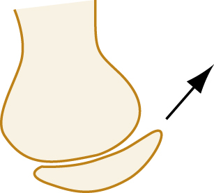
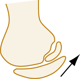
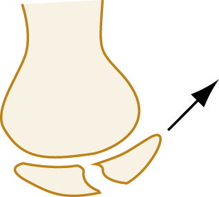
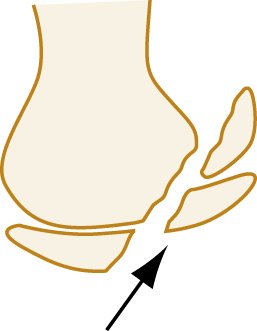
The term avulsion fracture refers to a bone fragment that is pulled away from its normal position by the forceful contraction of a muscle ( Fig. 41.5A ) or the resistance of a tendon or ligament to a force in the opposite direction (see Fig. 41.5B ). The term impaction refers to the forceful collapse of one fragment of bone into or onto another. In the proximal humerus, this collapse typically occurs in a telescoping manner, particularly in older patients, whose bones are osteoporotic and brittle. In the tibial plateau, impaction occurs frequently in the form of a depression ( Fig. 41.6A and B ) and, in the vertebral bodies, impaction frequently occurs in the form of compression resulting in a significant loss of bone height in some cases (see Fig. 41.6C ).
Avulsion Fractures.
(A) Musculotendinous avulsions of small bone fragments from the head of the humerus (arrows) . (B) Extensor tendon avulsion of bone from the base of the middle phalanx (arrow) .
(A and B) Tibial plateau fracture. (C) Vertebral body compression fracture (arrows) .
A fracture that occurs through abnormal or diseased bone is termed pathologic. A pathologic fracture is suggested whenever a fracture occurs from seemingly trivial trauma. Diseases that cause structural weakness predisposing to injury include primary malignancy or malignant metastatic lesions, bone cysts, enchondromas, and giant cell tumors. In addition, metabolic and genetic disorders such as osteomalacia, scurvy, rickets, vitamin D deficiency, Paget disease, and osteogenesis imperfecta can alter bone density, making them susceptible to fracture. The term pathologic is also often applied to fractures through osteopenic bone when demineralization is a result of disuse, as in polio. In contrast, fractures through osteoporotic bone of older adults usually are not described as pathologic; these are more accurately referred to as geriatric or insufficiency fractures. When fractures occur in normal bones and a history of supposed trivial trauma or a suspicious mechanism is elicited, interpersonal violence or abuse should be suspected, and safety of the patient assured.
Repeated low-intensity forces may lead to resorption of normal bone, resulting in a stress fracture. Other terms for this condition are fatigue fracture and march fracture (see Table 41.1 ). Most stress fractures occur in the lower extremities and affect individuals involved in activities such as long-distance running, basketball, aerobics, and dancing. There is often a history of functional pain leading up to the fracture. Extrinsic factors such as training regimens, type of equipment used, nutrition habits, as well as intrinsic factors such as anatomic variation, muscle endurance, and hormonal factors, have all been associated with stress fractures. The tibia, fibula, metatarsals, navicular, cuneiform, calcaneus, femoral neck, or femoral shaft may be involved. These injuries may not be recognizable on initial plain films. Management therefore should be based on the clinical diagnosis.
Fracture Eponyms and Mechanistic Names
Many fractures were described before the advent of radiography and are described shorthand by an eponym or other name rather than by the exact bony injury. These eponyms and mechanistic names reflect the storied history of medicine and orthopedic care and are still commonly used to describe specific orthopedic injuries (see Table 41.1 ).
Epiphyseal Fractures
Fractures involving the epiphyses in children and adolescents are described according to the Salter-Harris classification ( Table 41.2 ). The clinical features of fractures in children are further discussed in Chapter 170 .
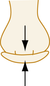
TABLE 41.2
Salter-Harris Classification
| Type | Description | Diagram |
|---|---|---|
| I | Fracture extends through the epiphyseal plate, resulting in displacement of the epiphysis; this may appear merely as widening of the radiolucent area representing the growth plate. |
|
| II | As above; in addition, a triangular segment of metaphysis is fractured. |
|
| III | Fracture line runs from the joint surface through epiphyseal plate and epiphysis. |
|
| IV | Fracture line occurs as in type III but also passes through adjacent metaphysis. |
|
| V | This is a crush injury of the epiphysis; it may be difficult to determine by radiographic examination. |
|
Clinical Features of Fractures
Fracture Healing
The goal of fracture reduction is to realign major bony fragments so that union can take place, and normal function is restored. In the initial stage of healing, a hematoma caused by the rupture of a vessel crossing the fracture line forms a hematoma. This hematoma eventually resorbs and provides the first continuity between the fragments. This procallus provides no structural rigidity for bearing stress but, with calcification and remodeling, callus is formed on the periosteal and endosteal surfaces of the bone, acting as a biologic splint. Over several months to a year, the callus normally completely ossifies, remodels, and becomes indistinguishable from mature bone.
Radiographic studies conducted 10 to 14 days after injury further reveal the fracture line as it becomes more visible due to localized bone resorption and hyperemia during the inflammatory phase. After 2 to 4 weeks, soft tissue swelling has regressed, and callus first becomes visible, initially in a mottled pattern and then taking on a dense appearance. Callus then undergoes organization, with peripheral margins becoming smooth as physically unstressed portions are resorbed.
In a healthy adult, the entire process from injury to consolidation takes approximately 2 months for the humerus and about 4 months for a large bone such as the femur. The rate of fracture healing is affected by the type of bone (cancellous bone heals faster than cortical bone), degree of fracture and opposition, systemic states, such as hyperthyroidism, use of corticosteroids, smoking, or illness requiring immobilization. Oblique fractures tend to heal more quickly than transverse fractures because of the greater amount of surface contact and a buttressing effect. Appropriately timed weightbearing can increase the rate of ossification of callus, whereas premature or excessive weightbearing can create a nonunion.
The presence of abundant bridging callus that is beginning to organize radiographically is usually associated with clinical union. If there is any suggestion of movement at the fracture site noted on clinical examination, union is regarded as inadequate. Several terms are used to denote abnormal healing. Delayed union is union that takes longer than usual for a particular fracture location; malunion occurs when a residual deformity exists; and nonunion is the failure of a fracture to unite. When nonunion results in a false joint, it is termed a pseudarthrosis.
If there is clinical evidence of stability, such as pain-free weightbearing, and radiographs demonstrate bridging bone at the cortices, a patient may resume activities of daily living with the injured extremity, even if the original fracture remains visible. The process of complete radiographic consolidation or healing can take several additional months.
Complications of Fractures
Infection (Osteomyelitis)
A fracture that communicates with the surface of the skin is termed an open fracture. Open fractures are treated as true, time-dependent orthopedic emergencies because of the risk of bone infection, or osteomyelitis . The high morbidity associated with osteomyelitis dictates that therapy be initiated expeditiously, including parenteral administration of antibiotics as early as possible, coverage with a moist dressing, and emergent washout of debris. The Gustilo-Anderson classification is commonly used to describe the various types of open fractures ( Box 41.2 ).
BOX 41.2
Classification and Emergency Management of Open Fractures
Grades
-
Grade I: Wound less than 1 cm long, punctured from below
-
Grade II: Laceration 5 cm long; no contamination or crush; no excessive soft tissue loss, flaps, or avulsion
-
Grade III: Large laceration, associated contamination or crush; frequently includes a segmental fracture
-
IIIA: Involves extensive soft tissue stripping of bone
-
IIIB: Periosteal stripping has occurred
-
IIIC: Major vascular injury present
-
Management
-
1.
Control hemorrhage in field with sterile pressure dressing after carefully removing gross debris (e.g., wood, clothing, leaves).
-
2.
Splint without reduction, unless vascular compromise is present.
-
3.
Irrigate with saline, cover with saline-soaked sponges after arrival in the emergency department.
-
4.
Begin IV antibiotic prophylaxis, usually a first-generation cephalosporin for grade I, with the addition of an aminoglycoside for grades II and III.
-
5.
Administration of tetanus prophylaxis, including tetanus immune globulin, for large crush wounds.
Suggested antibiotic therapy currently includes a first-generation cephalosporin, such as cefazolin, 2 g IV every 8 hours, for all open fractures, with the addition of an aminoglycoside, such as gentamicin, 5 mg/kg once daily, for type II or III fractures. Alternatively, a beta-lactam with both gram positive and negative coverage (e.g., ceftriaxone or piperacillin-tazobactam) may provide equivalent benefit. For patients with farm-related injuries or those with potential fecal contamination, ampicillin or penicillin should be added to the treatment regimen to empirically cover for clostridial contamination. Early versus delayed debridement of open fractures and the subsequent effect on rates of infection has been a source of debate. Historic guidelines recommending debridement of open fracture wounds within 6 hours of injury were based on animal experiments conducted in the 1890s. The timing of debridement—less than 6 hours versus more than 6 hours after injury—has not been proven to change outcome, but general practice is to undertake debridement and irrigation of the wound within the first 24 hours of injury. Regardless, the goals of open fracture management in the ED should focus on early administration of antibiotics, tetanus prophylaxis, dressing coverage of the wound, and splinting of the extremity.
Open distal tuft fractures of the fingers and toes are a notable exception to the previous recommendation. These are common when the phalanx of a finger or toe is subject to crush injury (e.g., by a closing door) and there is a skin wound overlying a fractured bone. Treatment of these injuries can be provided by the emergency clinician without the need for immediate consultation. Vigorous irrigation and debridement are adequate primary treatment for these injuries, provided digital arteries are intact. Infections of the tuft region are rare.
Hemorrhage
Because of the rich blood supply to the skeleton, fractures can result in large amounts of blood loss, shock, and even death from exsanguination. In particular, certain pelvic fractures can cause major blood loss because adequate tamponade is not possible. In adults, blood loss can range from 100 mL from a forearm fracture to 3 L from a pelvic fracture ( Table 41.3 ). Such hemorrhage can be controlled in part by early stabilization of the injured area through splinting, a binder, or skeletal traction. Definitive treatment options include embolization by an interventional radiologist or by emergent surgical intervention (see Chapter 46 ).
TABLE 41.3
Blood Loss Associated With Fracture in Adults
| Fracture Site | Amount of Blood Loss (mL) |
|---|---|
| Radius and ulna | 150–250 |
| Humerus | 250 |
| Tibia and fibula | 500 |
| Femur | 1000 |
| Pelvis | 1500–3000 |
Vascular Injuries
Vascular injuries are characteristically associated with certain fractures and may be limb threatening. Fractures and dislocations at the femorotibial articulation of the knee result from tremendous force, which may injure the popliteal artery and lead to the need for amputation (see Chapter 48 ). Fracture of the femoral neck requires emergent fixation regardless of the need for reduction to protect the blood supply to the femoral head and prevent aseptic necrosis. In the extremities, assessment of vascular injuries may be challenging. The initial survey should note the presence or absence of pulses and the state of capillary filling. If an end artery is completely disrupted, the tissue distal to the injury may exhibit the classic five Ps: pain and paresthesias (in the conscious patient), followed by pallor, pulselessness, and paralysis. Incomplete and subclinical injuries occasionally occur that initially may be asymptomatic and undetectable. In an unconscious, multiple trauma injured patient, major vascular injuries may not be obvious and may be overlooked. The mechanism of injury and anatomy dictate the need to assess the possibility of an injured vessel. If pulses cannot be palpated, a Doppler stethoscope should be used to detect blood flow. Even the presence of pulses may be misleading however, because pulses may be normal in some patients with significant arterial injuries. When pulses are present, but the mechanism of injury suggests the possibility of a vascular injury, additional diagnostic studies or surgical exploration may be necessary. If a limb is clearly not perfused, operative vascular exploration and repair should be undertaken promptly. Late complications of undiagnosed vascular injuries include thrombosis, arteriovenous fistulae, aneurysm, false aneurysm, and tissue ischemia with limb dysfunction. Delay of vascular injury repair is a risk factor for consequent amputation. The evaluation of peripheral vascular injuries is further discussed in Chapter 40 .
Related posts:


Full access? Get Clinical Tree


