CHAPTER 18. Abdominal Trauma
Reneé Semonin Holleran
COMPETENCIES


Get Clinical Tree app for offline access
1. Perform an organized and focused abdominal assessment.
2. Identify clinical indications of abdominal trauma.
3. Initiate critical interventions and provide appropriate treatment during transport for the patient with an abdominal injury.
Regionalized trauma care has drastically reduced the incidence rate of death after injury. However, despite improvements in prehospital, resuscitative, surgical, and critical care, unrecognized abdominal injury remains a preventable cause of death after injury. 2,3,15
Exsanguination continues to be a common cause of death from traumatic injury. Because patients with abdominal trauma may have severe hemorrhage, rapid transport can significantly reduce the mortality and morbidity rates from both blunt and penetrating abdominal injuries. Four common causes of massive bleeding in trauma patients include external injury, massive hemothorax, retroperitoneal injury (e.g., pelvic fracture, renal laceration, or major vessel lesion), and intraperitoneal injury (e.g., liver, spleen, or major vessel laceration). 3,9,15,17 The likelihood of significant intraabdominal injury is high when a patient has hypotension in the field, a major chest injury, or pelvic fracture. 14,35,42
Patients with genitourinary injuries alone are not usually in danger of life-threatening hemorrhage. Although genitourinary trauma alone is not immediately life threatening, appropriate transport can significantly affect the recovery time and reduction of complications that result from these injuries. Many lower level facilities are not equipped to diagnose and treat specific genitourinary injuries. Air medical transport may be appropriate when a delay in ground time diminishes the chances of full recovery from renal, bladder, and genital injuries.
Shock and time elapsed between injury and arri-val in the operating room are the major factors that affect survival of patients with abdominal injuries, and these factors can be ameliorated by intervention of trained transport personnel able to recognize life-threatening injuries, initiate treatment, and rapidly transport the patient to definitive care.
ABDOMINAL TRAUMA
Anatomy of the Abdomen
The abdomen contains several major organs of the body responsible for digestion, nutrition, and elimination of toxins and waste from the body. Because the spleen filters aged red blood cells and one of the functions of the liver is to eliminate toxic waste from the bloodstream, both organs are vascular in nature. The spleen, liver, and vascular system of the abdomen are the primary sources of exsanguination during abdominal trauma. 1,2,11,18,19 Injuries to the hollow abdominal organs, such as the small and large intestine, can result in abscess formation, wound infection, and sepsis, especially if trauma to the intestine remains undiagnosed for a period of time. When injuries to the abdomen occur, they may have a large impact on morbidity and mortality.
The abdominal cavity includes all structures and organs between the respiratory diaphragm and the urogenital diaphragm. This space is divided into three compartments: the first, and largest, is the peritoneal cavity; the second is the space within the pelvic structure; and the third is the retroperitoneal space. The peritoneal cavity contains the diaphragm, spleen, liver, stomach, transverse colon, and most of the small intestine and mesentery.
The bony structure of the pelvis contains the rectum, bladder, iliac vessels, and female reproductive organs; the penis and scrotum are located outside the abdominal cavity below the urogenital diaphragm.
The retroperitoneal space is separated from the abdominal cavity by the posterior peritoneum and therefore is not always accessed with peritoneal lavage. Injuries in this area may be difficult to diagnose and are easily overlooked. The organs contained in this space are the aorta, vena cava, distal esophagus, kidneys, ureters, and portions of the duodenum, pancreas, colon, and rectum.
Knowledge of the basic anatomy of the abdomen and position of the organs is crucial because identification of organs possibly injured by blunt or penetrating trauma helps the transport team provide the treatment necessary for those injuries when preparing for or affecting the patient’s transport.
Classification of Injuries
Abdominal trauma can be blunt or penetrating, depending on the mechanism of injury. Blunt trauma is caused by any type of force being exerted on the abdomen as the result of falls, motor vehicle accidents (MVCs), bicycle and motorcycle accidents, or any force striking the abdomen. 1,4,7,8,12,28 As the body receives an impact, the organs in the abdominal cavity continue moving forward; vessels and tissues tear away from their attachment points.
Patients have fewer fatal injuries with the increased use of seat belts, motorcycle and bicycle helmets, child restraint seats, and air bags and with enforcement of alcohol restraints. However, most blunt abdominal injuries (50% to 75%) are the result of MVCs. 1,7,11,13 The use of seat belts decreases the possibility of multisystem injuries, but improper placement across the abdomen rather than the pelvis, loose application, or use of only lap belts can result in visceral trauma. The abdominal organs most frequently injured by blunt trauma are the liver, spleen, and kidney, although hollow-organ (intestinal) injury can occur. Abdominal injuries among air bag–protected occupants occur less frequently than head, chest, and lower extremity injuries. However, abdominal injuries may be occult, and deformation of the steering wheel is an indicator of an increased likelihood of internal injury. 3,4,23,44.45.48.47. and 48.
Trauma is classified as penetrating when an object such as a knife or bullet enters the abdominal cavity; anything that has penetrated the abdomen and remains in place may be designated as an impaled object. Penetrating injuries of the abdomen are caused by gunshot wounds (GSWs) or stab wounds. The degree of injury of patients with GSWs depends on the caliber of the gun and its distance from the patient. High-velocity weapons and close-range shotguns cause more destruction to abdominal organs than do low-velocity weapons. However, bullets from low-velocity weapons can deflect off organs and bony prominences and create extensive injuries that are not easily recognized on initial examination. In stab wounds, the length of the knife or object, the depth of penetration, and the angle at which it was inserted determine the amount of injury. The major organs involved in penetrating injuries are the liver, small bowel, colon, stomach, and vascular structures. The large size and anterior position of the liver and bowel in the abdomen make them particularly vulnerable to penetrating trauma. 12,21.22.23.24. and 25.
Objects found impaled in the patient on arrival of the transport team should be left in place and stabilized for transport (Figure 18-1). Removal of the object may cause further injury or increase bleeding. If placing the patient into the transport vehicle is not possible because of the size and position of the object, it may have to be cut off while still in place within the abdominal cavity. The object should be moved as little as possible. In some instances, the trauma surgeon must be transported to the scene to assist in the shortening or removal of the object. 41
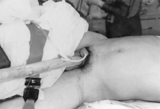 |
| FIGURE 18-1 Injury resulting from a logging accident. |
Patient History
The history of the injury and the physical examination should be obtained before transport because the information is important to the transport team when they assess and stabilize a patient for transport. The trauma surgeon may use the information when deciding the degree of injury and whether surgery should be performed soon after arrival at the emergency center. When possible, a medical history, including medications, allergies, illnesses, and events leading up to injury, should be obtained from the patient, family, or referring facility. Other helpful information is whether the patient has used alcohol or other substances, has a head or neck injury, has psychiatric problems, or has any underlying medical conditions (i.e., cardiovascular disease or coagulopathies). 1,10,15 Initial vital signs and level of consciousness, intake and output, and treatments done before the arrival of the transport team, and any changes or treatments in transport, should be reported to the trauma team at the receiving trauma center.
When a patient with blunt abdominal trauma is transported from a pedestrian accident or MVC, important information to obtain from the prehospital personnel at the site are the time of injury, probable speed of impact, damage to the vehicle (steering wheel, direction of impact on the vehicle), patient’s position in the vehicle, and restraint devices used. When time permits, brief inspection of the damage to the vehicle by the transport team may provide more information on possible patient injuries. In patients injured from falls, the height of fall and position of impact help point to the type of injuries involved. The history of assault victims should include the type of instrument that struck them. 24,26
Although internal bleeding is difficult to measure in penetrating injuries, the amount of blood lost at the scene should be noted for determination of the fluid replacement needed and indication of the degree of injury.
Physical Examination
The physical examination is one of the most important procedures for diagnosis of abdominal injury. It can be effective in determining the presence of blood in the peritoneal cavity or peritoneal irritation. Examination of the abdomen should be as thorough as possible. When a trauma patient who is in hemorrhagic shock is transported from a scene of accident or assault, the only assessment that time may permit is palpation for distention and tenderness. The abdominal assessment should include inspection, auscultation, and palpation before interfacility transport, especially when time and distance are great. Serial abdominal assessments should be done throughout transport because peritoneal irritation and accumulation of blood in the peritoneal cavity may not produce symptoms immediately. 1
The abdomen should be fully exposed to allow the transport team to inspect for contusions, abrasions, deformity, hematoma formation, open wounds, and penetrating injuries. An ecchymotic discoloration around the umbilicus (Cullen’s sign) can indicate intraabdominal or retroperitoneal hemorrhage. When examination of the patient’s back is possible, the transport team should look for any obvious signs of trauma that indicate an abdominal or genitourinary injury. Flank bruising (also known as Grey-Turner’s sign) may signify retroperitoneal hematoma formation from trauma to the kidneys, major blood vessels, or other organs contained in that space.
Auscultation should be done primarily in a quiet controlled area because bowel sounds are difficult to hear in the noisy transport environment. Absence of bowel sounds can be an important indicator of an ileus and abdominal injury, and assessment of bowel sounds before an interfacility transfer may alert the transport team to the possibility of abdominal injury. Absence of bowel sounds may indicate the need to consider the insertion of a gastric tube for patient comfort and to protect the airway from potential aspiration.
An abdomen that is tender and distended on palpation needs immediate attention, which generally requires rapid transport with continual observation of vital signs. The major causes of a distended abdomen are gastric dilation and rapid intraabdominal bleeding. Tenderness on palpation with involuntary guarding or rebound tenderness is indicative of peritoneal irritation.
Subjective reports of abdominal pain by a trauma patient should always be addressed. Because patients with head injury and a decreased level of consciousness are unable to identify abdominal pain, detection of a distended abdomen on palpation without any obvious signs of trauma may be the only indication of intraabdominal trauma.
The perineum should be visually inspected for any injuries to the genitals, urethra, or rectum. Peroneal hematoma formation can be caused by a retroperitoneal hematoma, pelvic fracture, or direct peritoneal trauma.
Diagnostic Procedures
Three diagnostic studies are used to evaluate the injured abdomen. These studies are diagnostic peritoneal lavage (DPL), ultrasound scan (focused abdominal sonogram for trauma), and computerized axial tomographic (CT) scan. 1,7,19,20,30,34.35.36.37. and 38.
Diagnostic peritoneal lavage was the standard method used to evaluate abdominal trauma for decades. However, the advent of newer, more accurate, and less invasive technologies has made this procedure less commonly used than it once was. The American College of Surgeons Committee on Trauma recommends that a DPL be performed in a patient with an unstable condition if the abdominal examination results are suggestive of injury or unreliable. 1,2,19
Focused abdominal sonogram for trauma (FAST) is generally used for torso evaluation of patients with multiple traumas. 28 The FAST examination is rapid and noninvasive but needs to be performed by qualified persons. It also can be distorted by bowel, gas, and subcutaneous air. It does not detect all abdominal injuries. Because intraabdominal injury cannot be totally ruled out with negative FAST results, especially in unstable conditions, a computed tomographic (CT) scan of the abdomen should be performed. 7,28
Computed tomography of the abdomen for both adults and children offers both high specificity and sensitivity to the detection and location of abdominal injury. CT scan is effective in demonstrating solid viscous injuries such as spleen or liver lacerations, the presence and quantity of hemoperitoneum, and retroperitoneal injuries such as renal lacerations or hematomas associated with pelvic fractures. The CT scan has been helpful in decreasing the number of unnecessary laparotomies. 15,24,28
Use of Ultrasound Scan Before and During Transport
In the past, several factors presented challenges to the use of ultrasound technology in the field. These factors included the size of the machine, the fragility of ultrasound probes, and the skills of those performing the tests. However, the availability of new technology has made it possible to use ultrasound in the transport environment. 30,36,38,43 Price, Wilson, and Murphy, 43 Mazur et al, 36 Melanson et al, 38 and Knobloch30 have all looked at the advantages and disadvantages of performing a FAST examination before and during transport. Advantages include early identification of intraabdominal free fluid in the prehospital environment, which can contribute to rapid diagnosing of potentially life-threatening abdominal injuries and assure that the patient is transported to the most appropriate center for care. Another use of ultrasound scan in the prehospital environment is the evaluation of the ventricular situation before and after CPR, which could prevent the unnecessary transport of a dead patient. 30
The disadvantages of the use of ultrasound scan in the transport environment have included the limited ability of transport team members to adequately perform the examination, the small size of the screen, the brightness of the screen, the cramped space within the aircraft and limited flight times, and the overall fragility of the equipment. 38 As technology continues to develop, a FAST examination before or during transport may become a useful assessment tool for the patient with abdominal trauma.
Patient Assessment, Treatment, and Transport
In many cases, injury is not limited to the abdomen, particularly in patients with blunt abdominal trauma. Once the primary assessment is completed and critical interventions initiated, the secondary examination should be done for determination of any other injuries.
In addition to the usual airway and breathing assessment and management, signs and symptoms of cardiovascular system collapse, such as hypotension, delayed capillary refill, and decreased level of consciousness, are important indicators of abdominal injuries. Patients with visceral injuries have the potential for sudden onset of severe hypovolemic shock because of the vascular nature of many organs in the abdominal cavity and the space for occult blood accumulation. Rapid recognition of the possibility of major abdominal injury and rapid transport after the initiation of critical interventions should be the goal when a patient is in hemorrhagic shock or has impending exsanguination.
Patients in severe shock or near exsanguination exhibit a decreasing level of consciousness as hypoxia increases and pale to mottled skin that is cold, clammy, and possibly profusely diaphoretic (Box 18-1). Mucous membranes may be pasty white, and capillary refill is delayed or absent. When no obvious external bleeding is present, the transport nurse should examine the abdomen. If the abdomen is distended and rigid with no bowel sounds, the transport team member should suspect intraabdominal bleeding. The conscious patient reports severe abdominal pain. If the abdomen is tautly distended, pressure is exerted on the diaphragm, causing potential shortness of breath and tachypnea. The patient is tachycardic and hypotensive; a blood pressure reading may not be obtainable even with a weak brachial pulse. If the blood pressure does not respond to fluid administration, exsanguination is a real possibility. The survival of a patient who is exsanguinating may depend on the amount of time it takes for transport to definitive care. Not all patients with abdominal injuries display all the symptoms described previously. Varying degrees of these symptoms, such as abdominal tenderness without initial distention, decreased but not absent bowel sounds, and tachycardia without hypotension, should alert the transport team to the possibility of exsanguination.
BOX 18-1
Symptoms of Shock
Decreasing consciousness
Pale-to-mottled skin that is cold, clammy, and possibly diaphoretic
Pasty white mucous membranes
Delayed or absent capillary refill
Distended and rigid abdomen with no bowel sounds
Potential shortness of breath and tachypnea
Tachycardia and hypotension
Unobtainable blood pressure reading
Blood pressure that does not respond to fluid administration
Reports of severe abdominal pain
Initial management at the scene when transport time should be short should consist of airway management, 100% oxygen delivery, and spinal protection. For the patient with an abdominal injury, fluid resuscitation should be initiated. This should be done while constantly monitoring the patient during transport. In addition to crystalloids, blood and blood products may need to be administered. The likelihood of significant intraabdominal injury is high when a patient has hypotension in the field, a major chest injury, or pelvic fracture.
Interfacility transfers may increase the patient transport time. Patient management should include all the treatment instituted for the short-scene-call transport. In addition, a gastric tube should be inserted for suspected intestinal injury, gastric distention, or aspiration. If massive external or internal hemorrhage is occurring, the referring hospital may have type-specific or O negative blood infusing or ready to send with the patient. If the report before transport indicates hypovolemic shock, the transport team should request that the referring hospital have blood ready for transport on the patient’s arrival or obtain blood products for use during the transport. For some patients, blood transfusion before arrival at the level I trauma center can be the factor that decreases morbidity and mortality. Hypothermia caused by massive intravenous fluid resuscitation can have a negative influence on patient outcome; therefore, warming of intravenous fluids for the administration of many liters of fluid, or at least use of fluids from an emergency department instead of cold fluids in the transport vehicle, can be important. The goals of transport are initiation of critical interventions and rapid transport to the nearest facility capable of treating the injured patient. 5,6,14,29,32,40,45
SPECIFIC ABDOMINAL ORGAN INJURIES
Diaphragm
Incidence and Mechanism of Injury
Blunt injury to the diaphragm, resulting in rupture or partial tear, occurs when a tremendous force is applied to the abdomen. The left diaphragm is injured more often than the right because the liver absorbs the impact of the force on the right side. If a right-sided tear has occurred, liver injury will probably accompany it. Spleen injuries often occur with left-sided diaphragmatic trauma. 1,2,15,19,20 Diaphragmatic tears can occur without herniation of the bowel into the chest cavity. If an intestinal herniation into the pleural space does occur, intestinal strangulation may develop. A penetrating injury of the diaphragm should be suspected when a knife wound occurs at or below the nipple line anteriorly or at the inferior border of the scapula posteriorly. 2,50
Assessment and Symptoms
The transport team may not be able to diagnose a diaphragmatic tear that has not resulted in bowel herniation. Further diagnostic procedures may be necessary to ascertain a diaphragmatic tear. Physical examination of the patient with a diaphragmatic hernia may reveal absent or reduced breath sounds on the affected side. Bowel sounds may be heard in the chest cavity when intestinal contents have herniated into the pleural space. Respiratory distress may accompany intestinal herniation. If stomach contents are returned when a needle thoracostomy is performed for a believed tension pneumothorax, diaphragmatic hernia should be suspected. Chest radiographic results may indicate intestinal herniation with the presence of stomach contents or a curled gastric tube in the chest cavity. Diaphragm injuries missed on diagnostic examination may result in intestinal incarceration or strangulation that occurs weeks to months later. Associated mortality rates are high. 2
< div class='tao-gold-member'>
Only gold members can continue reading. Log In or Register to continue
Related posts:


Full access? Get Clinical Tree
