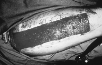
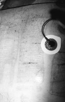
PROCEDURE 119 • A partial-thickness wound is surgically created when a donor site (Fig. 119-1) is harvested to obtain skin for a full-thickness defect. The more dermis moved with the skin graft, the less the graft shrinks with healing; therefore, deeper donor sites may be created to obtain skin for cosmetically significant areas such as the face or hands.9 Depending on the percentage of dermis moved with the graft, donor sites created may be superficial or deep partial-thickness wounds that heal in 10 to 20 days (typically, 10 to 14 days; Fig. 119-2).6 • Factors that can disrupt or prolong healing include infection, desiccation, edema, adherent dressing changes, poor nutrition, hemodynamic instability, and a variety of preexisting medical conditions.2 • The longer a partial-thickness wound takes to heal, the more significant the scarring; therefore, donor sites can produce minimal or hypertrophic scars.2 Donor sites retain deep epidermal appendages, so they are generally capable of sweating and bearing hair after they heal. The donor site may be reharvested once healing is complete, but skin from the first harvest of a donor site is always of higher quality than that of repeat harvesting. • Because the dermis is richly supplied with capillaries and nerve endings, donor sites are at risk for bleeding in the first 24 hours and are exquisitely tender to touch. They produce large volumes of serous exudate. • Donor site treatment goals include minimizing bleeding, supporting reepithelialization, managing exudate, preventing infection, controlling pain, and minimizing scarring.9 Epinephrine-soaked dressings, thrombin spray, or compression dressings may be applied in the operating room to attain stasis.9 A compression dressing is usually used for the first 12 to 24 hours to ensure stasis.8 After this initial period, compression may be applied for comfort. • Wounds epithelialize most rapidly in a moist environment. If donor sites are small enough, use of a thin-film polyurethane or hydrocolloid dressing has been shown to promote rapid healing while providing comfort through dressing flexibility and occlusive coverage of nerve endings.6 Occlusive dressings (sealed on all sides) can be difficult to maintain on larger donor sites because of the substantial volume of exudate.6 A small drain (attached to a vacutainer and collection tube Becton, Dickinson and Co., Franklin Lakes, NJ) tube can be used to remove excess drainage from under a thin-film dressing. Calcium alginates have also been used successfully under occlusive dressings to mange exudate.6 • Multilayer occlusive dressings may also be used, with a nonstick dressing (e.g., greasy gauze, meshed silicone) applied next to the donor site and a bulky absorbent outer layer to maintain a moist environment and wick away excess drainage. The outer dressing may be changed periodically, leaving the inner dressing intact until the wound heals beneath it. • One of the oldest and most cost-effective methods for treatment of donor sites is to apply mesh gauze, wrap with an outer wrap for 12 to 24 hours, and then remove the outer wrap and allow the inner dressing to remain exposed and dry until the wound heals beneath. This method has been done with fine mesh gauze and xeroform (McKesson Brand, San Francisco, CA)/xeroflo (Kendall Healthcare, Coviden, Mansfield, MA). The technique is only effective if the dressing dries well and becomes impermeable to bacteria, essentially acting as a scab.11 Positioning the patient for maximal exposure of the donor sites, preventing prolonged donor site contact with sheets and clothing, and increasing airflow across the wound are important for this technique to work.5 If the donor site is large, this procedure creates a rather stiff and uncomfortable protective layer. • Biobrane (UDL Laboratories, Rockford, IL), a biosynthetic product, produces a more flexible donor site dressing. When exposed to air at 24 hours after harvesting, it dries to form a fibrous bond with the collagen in the wound. This dressing provides improved pain control, exudate management, and rate of healing when compared with a fine-mesh gauze dressing.12 • Antimicrobial creams or ointments have also been used on donor sites, essentially treating the wounds in the same way as partial-thickness burns are treated. The disadvantage to this approach is that it requires daily washing of the wound and reapplication of cream and dressings.11 • Slow-release silver dressings are gaining popularity for donor site use. These dressings are moistened twice daily with sterile water to release the silver. In highly exudating wounds, the wound moisture may be adequate to promote silver release without exogenously applied moisture. These dressings release silver for 3 or more days; ideally, they are placed on the donor site in the operating room, and the wound is allowed to heal beneath with infrequent or no dressing changes.6,11 • Donor sites need to be assessed daily for signs of infection, including periwound warmth and erythema, increased pain, and purulent drainage. Bacteria can delay healing and increase scarring or convert a partial-thickness donor site to a full-thickness wound.2
Donor Site Care
PREREQUISITE NURSING KNOWLEDGE
Related posts:
![]() 39: Automated External Defibrillation
39: Automated External Defibrillation
![]() 139: Calculating Doses and Flow Rates and Administering Continuous Intravenous Infusions
139: Calculating Doses and Flow Rates and Administering Continuous Intravenous Infusions
![]() 16: Continuous Venous Oxygen Saturation Monitoring
16: Continuous Venous Oxygen Saturation Monitoring
![]() 132: Small-Bore Feeding Tube Insertion Using an Electromagnetic Guidance System (CORTRAK®)
132: Small-Bore Feeding Tube Insertion Using an Electromagnetic Guidance System (CORTRAK®)
![]() 83: Implantable Venous Access Device: Access, Deaccess, and Care
83: Implantable Venous Access Device: Access, Deaccess, and Care
![]() 64: Blood Sampling from an Arterial Catheter
64: Blood Sampling from an Arterial Catheter

Full access? Get Clinical Tree


119: Donor Site Care